Skip to main page content
U.S. flag

An official website of the United States government

Dot gov

The . gov means it’s official. Federal government websites often end in . gov or . mil. Before sharing sensitive information, make sure you’re on a federal government site VSports app下载. .

Https

The site is secure. The https:// ensures that you are connecting to the official website and that any information you provide is encrypted and transmitted securely. V体育官网.

. 2009 Aug 19;4(8):e6691.
doi: 10.1371/journal.pone.0006691.

Multi-modality therapeutics with potent anti-tumor effects: photochemical internalization enhances delivery of the fusion toxin scFvMEL/rGel

Affiliations

Multi-modality therapeutics with potent anti-tumor effects: photochemical internalization enhances delivery of the fusion toxin scFvMEL/rGel

Pål K Selbo et al. PLoS One. .

Abstract

Background: There is a need for drug delivery systems (DDS) that can enhance cytosolic delivery of anti-cancer drugs trapped in the endo-lysosomal compartments. Exposure of cells to specific photosensitizers followed by light exposure (photochemical internalization, PCI) results in transfer of agents from the endocytic compartment into the cytosol VSports手机版. .

Methodology and principal findings: The recombinant single-chain fusion construct scFvMEL/rGel is composed of an antibody targeting the progenitor marker HMW-MAA/NG2/MGP/gp240 and the highly effective toxin gelonin (rGel). Here we demonstrate enhanced tumor cell selectivity, cytosolic delivery and anti-tumor activity by applying PCI of scFvMEL/rGel. PCI performed by light activation of cells co-incubated with scFvMEL/rGel and the endo-lysosomal targeting photosensitizers AlPcS(2a) or TPPS(2a) resulted in enhanced cytotoxic effects against antigen-positive cell lines, while no differences in cytotoxicity between the scFvMEL/rGel and rGel were observed in antigen-negative cells. Mice bearing well-developed melanoma (A-375) xenografts (50-100 mm(3)) were treated with PCI of scFvMEL/rGel V体育安卓版. By 30 days after injection, approximately 100% of mice in the control groups had tumors>800 mm(3). In contrast, by day 40, 50% of mice in the PCI of scFvMEL/rGel combination group had tumors<800 mm(3) with no increase in tumor size up to 110 days. PCI of scFvMEL/rGel resulted in a synergistic effect (p<0. 05) and complete regression (CR) in 33% of tumor-bearing mice (n = 12). .

Conclusions/significance: This is a unique demonstration that a non-invasive multi-modality approach combining a recombinant, targeted therapeutic such as scFvMEL/rGel and PCI act in concert to provide potent in vivo efficacy without sacrificing selectivity or enhancing toxicity. The present DDS warrants further evaluation of its clinical potential V体育ios版. .


Conflict of interest statement

Competing Interests: The authors have declared that no competing interests exist.

Figures

Figure 1
Figure 1. Photochemical-mediated endo-lysosomal release and cytosolic delivery of photosensitizer and scFvMEL/rGel.
A375-GFP cells were incubated for 18 h with 5 µg/ml AlPcS2a, washed twice and chased in drug-free medium for 4 h. 30 min prior to microscopy, cells were incubated with 5 µg/ml Hoechst 33342. A, Fluorescence of EGFP, Hoechst 33342 and AlPcS2a and merged (E+H+A) in vital cells prior to (upper panels) and 1 min post 10 sec of microscopy red light exposure (590–650 nm) with respectively merged micrographs and corresponding phase contrast (mid panels). Structure formula of AlPcS2a in left lower panel. B, A-375 cells were co-incubated for 18 h with 80 nM scFvMEL/rGel and 5 µg/ml AlPcS2a, washed twice and chased in drug-free medium for 4 h prior to light exposure and further incubated at 37°C for 30 min prior to ICC detection of rGel. Right panel, negative controls included rabbit IgG isotype.
Figure 2
Figure 2. Enhancement of cytotoxicity and reduction of EGFP signals by PCI of scFvMEL/rGel in A375-GFP melanoma cells.
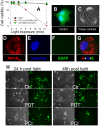
A, Viability of A375-GFP cells subjected to PCI of scFvMEL/rGel (red triangles), PDT (black dots) or scFvMEL/rGel only (green squares). Cells were incubated with 5 µg/ml AlPcS2a with or without 100 nM scFvMEL/rGel for 18 h, followed by 3 times wash and 4 h chase in drug-free medium prior to exposure to increasing light exposure of cells (15 minutes = 1.35 J/cm2). Cytotoxicity was assessed by the MTT assay 48 h post light exposure. Bars, SD. B, Fluorescence of EGFP and Hoechst 33342 in an untreated A375-GFP cell. C, Phase contrast of a cell treated with PCI of scFvMEL/rGel (LD90, 0.54 J/cm2) with corresponding fluorescence of AlPcS2a (D), Hoechst 33342 (E), EGFP (F) and a merge of D,E and F (G) 48 h post light exposure. H, A375-GFP cells 24 and 48 hours post either PDT (LD50, 1.35 J/cm2) or PCI of scFvMEL/rGel (LD90, 0.54 J/cm2) as compared to untreated control cells (Ctr).
Figure 3
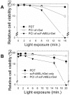
Figure 3. PCI of scFvMEL/rGel in MA11 breast carcinoma cells (A) and U87 malignant glioma cells (B).
The cells were incubated with 5.0 µg/ml AlPcS2a+/−16.5 nM scFvMEL/rGel or 16.5 nM rGel for 18 hours and then washed twice and chased in drug-free medium 4 h prior to red light exposure. MTT assay was performed 48 hours post light. Experiments with triplicates were reproduced at least twice. Bars, SE.
Figure 4
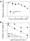
Figure 4. Control experiments in gp240-negative bladder cancer cell line T24.
A, Cells were treated with either rGel or scFvMEL/rGel for 18 h with increasing toxin concentrations. B, AlPcS2a-PCI of scFvMEL/rGel or rGel in T24 cells. The cells were treated and assessed as described in figure 3. Bars, SE.
Figure 5
Figure 5. PCI of scFvMEL/rGel in A-375 xenografts.
A, Kaplan Meier plot of treatment groups as indicated in the figure. Nude mice with s.c. growing A-375 (50–100 mm3) tumors were given AlPcS2a (5 mg/kg i.p.) with or without scFvMEL/rGel (2 mg/kg i.v) administered 48 and 24 hours prior to laser light exposure (day 0), respectively. The light dose was 20 J/cm2 with an irradiance of 100 mW/cm2. n, number of animals in each group. B, Weights of the mice treated with PCI of scFvMEL/rGel as compared with non-treated animals. Range of weights at treatment start were 22–29 g. Weight data for the other groups are shown in Figure S6.
Figure 6
Figure 6. Tumor response and skin regeneration post PCI of scFvMEL/rGel.
Two mice (#1 and #2) with ∼75 mm3 A-375 tumors at day 0 were further assessed at day 1, 5 and 19 post PCI. Both mice obtained CR and excellent wound repair after PCI of scFvMEL/rGel (still at day 110) as compared to a untreated control mouse (Ctr) having a tumor which reached >1000 mm3 at day 19 (∼13-fold tumor increase compared to day 0).

References

    1. Lu Y, Yang J, Sega E. Issues related to targeted delivery of proteins and peptides. AAPS J. 2006;8:E466–E478. - PMC - PubMed
    1. Berg K, Selbo PK, Prasmickaite L, Tjelle TE, Sandvig K, et al. Photochemical internalization: a novel technology for delivery of macromolecules into cytosol. Cancer Res. 1999;59:1180–1183. - PubMed
    1. Selbo PK, Sivam G, Fodstad O, Sandvig K, Berg K. In Vivo Documentation of Photochemical Internalization, a Novel Approach to Site Specific Cancer Therapy. Int J Cancer. 2001;92:761–766. - PubMed
    1. Hogset A, Prasmickaite L, Selbo PK, Hellum M, Engesaeter BO, et al. Photochemical internalisation in drug and gene delivery. Adv Drug Deliv Rev. 2004;56:95–115. - PubMed
    1. Berg K, Moan J. Lysosomes as photochemical targets. Int J Cancer. 1994;59:814–822. - PubMed

Publication types

MeSH terms

V体育平台登录 - LinkOut - more resources