Skip to main page content
U.S. flag

An official website of the United States government

Dot gov

The . gov means it’s official. Federal government websites often end in . gov or . mil. Before sharing sensitive information, make sure you’re on a federal government site. VSports app下载.

Https

The site is secure V体育官网. The https:// ensures that you are connecting to the official website and that any information you provide is encrypted and transmitted securely. .

. 2009 Aug 15;69(16):6668-75.
doi: 10.1158/0008-5472.CAN-09-1284.

Cullin 1 functions as a centrosomal suppressor of centriole multiplication by regulating polo-like kinase 4 protein levels

Affiliations

V体育安卓版 - Cullin 1 functions as a centrosomal suppressor of centriole multiplication by regulating polo-like kinase 4 protein levels

V体育ios版 - Nina Korzeniewski et al. Cancer Res. .

Abstract

Abnormal centrosome and centriole numbers are frequently detected in tumor cells where they can contribute to mitotic aberrations that cause chromosome missegregation and aneuploidy. The molecular mechanisms of centriole overduplication in malignant cells, however, are poorly characterized VSports手机版. Here, we show that the core SKP1-cullin-F-box component cullin 1 (CUL1) localizes to maternal centrioles and that CUL1 is critical for suppressing centriole overduplication through multiplication, a recently discovered mechanism whereby multiple daughter centrioles form concurrently at single maternal centrioles. We found that this activity of CUL1 involves the degradation of Polo-like kinase 4 (PLK4) at maternal centrioles. PLK4 is required for centriole duplication and strongly stimulates centriole multiplication when aberrantly expressed. We found that CUL1 is critical for the degradation of active PLK4 following deregulation of cyclin E/cyclin-dependent kinase 2 activity, as is frequently observed in human cancer cells, as well as for baseline PLK4 protein stability. Collectively, our results suggest that CUL1 may function as a tumor suppressor by regulating PLK4 protein levels and thereby restraining excessive daughter centriole formation at maternal centrioles. .


Figures
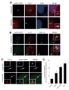
Figure 1
Figure 1. CUL1-positive maternal centrioles serve as assembly platforms for oncogene-induced centriole overduplication
(A) Immunofluorescence microscopic analysis for CUL1 using U-2 OS cells stably expressing centrin-GFP (U-2 OS/centrin-GFP). Arrowheads indicate centrioles shown in inserts. Nuclei stained with DAPI. Scale bar indicates 10 μm. (B) Co-immunofluorescence microscopic analysis of U-2 OS/centrin-GFP cells for CUL1 and CEP170, a marker for mature maternal centrioles. Arrowheads indicate centrioles with co-localization of CUL1 and CEP170 (see also inserts). (C) Immunofluorescence microscopic analysis of U-2 OS/centrin-GFP cells for CUL1 following overexpression of E2F-1. Note overduplication of centrioles in the presence of only two CUL1-positive centrioles (bottom panels). (D) Quantification of the proportion of U-2 OS/centrin-GFP cells with centriole overduplication in the presence of one or two CUL1-positive centrioles after overexpression of c-MYC, HPV-16 E7 or E2F-1. Arrows point to centrioles shown in inserts. Mean and standard error of three independent experiments with at least 100 cells counted per experiment are shown.
Figure 2
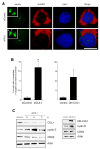
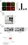
Figure 2. Inhibition of CUL1 causes centriole multiplication
(A) Fluorescence microscopic analysis of U-2 OS/centrin-GFP cells transfected with either control siRNA duplexes (siControl) or siRNA targeting CUL1 (siCUL1) for 72 h. A DsRED-encoding plasmid was used as transfection marker. Nuclei stained with DAPI. Arrowheads in bottom insert indicate centriole multiplication with three daughter centrioles at a single mother. Scale bar indicates 10 μm. (B) Quantification of centriole multiplication (>4 centrioles total, >1 daughter at a single mother) in U-2 OS/centrin-GFP cells transfected with either control (siControl) or CUL1 (siCUL1) siRNA duplexes for 72 h (left panel) or ectopic expression of empty vector control or DN-CUL1 for 48 h (right panel). Mean and standard error of three independent experiments with at least 100 cells counted per experiment are shown. Asterisk indicates statistically significant differences (p≤0.05; Student’s t test for independent samples). (C) Immunoblot analysis of whole cell extracts from U-2 OS/centrin-GFP cells transiently transfected with siRNA duplexes targeting CUL1 (siCUL1) or control siRNA (siControl) for the indicated time intervals (left panels) or ectopic expression of empty vector control or DN-CUL1 for 48 h (right panel). Immunoblots for CUL1, cyclin E and CDK2 are shown. DN-CUL1 was detected using an OctA-Probe/Flag® antibody (Santa Cruz). Immunoblot for Actin shows protein loading.
Figure 3
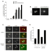
Figure 3. Ectopic expression of cyclin E/CDK2 does not stimulate centriole multiplication but causes
aberrant PLK4 recruitment to maternal centrioles. (A) Quantification of the proportion of cells with centriole multiplication (>4 centrioles, >1 daughter per maternal centriole) after transient transfection with empty vectors (controls) or either cyclin E/CDK2 alone (0 μg PLK4 plasmid DNA) or increasing amounts of PLK4 alone (grey bars) or a combination of cyclin E/CDK2 with increasing amounts of PLK4 plasmid DNA (black bars). Asterisks indicate statistically significant differences (p≤0.05 at 0.5 μg and p≤0.005 at 2 μg PLK4 plasmid DNA). (B) Fluorescence microscopic analysis of U-2 OS/centrin-GFP cells after transient transfection with empty vector (control) or cyclin E, CDK2 and PLK4. Note centriole multiplication in the right panel. (C) Immunofluorescence microscopic analysis of U-2 OS/centrin-GFP cells for endogenous PLK4 expression after transient transfection with either empty vector (control) or cyclin E/CDK2. Arrows indicate an aberrant daughter centriole that co-localizes with PLK4 at the maternal centriole. Arrowheads indicate an aberrant PLK4 signal at a maternal centriole without a detectable centrin-positive daughter. (D) Quantification of the percentage of U-2 OS/centrin-GFP or BJ/TERT/centrin-GFP cells with aberrant (two or more) PLK4 dots at maternal centriole following transfection with empty vector (control) or cyclin E/CDK2.
Figure 4
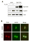
Figure 4. PLK4 is degraded by the proteasome
(A) Immunoblot analysis of whole cell extracts from U-2 OS/centrin-GFP cells transiently transfected with either empty vector (control) or PLK4 and treated with 1 μM of the proteasome inhibitor Z-L3VS or 0.1% DMSO at 24 h after transfection for an additional 24 h. (B) Immunofluorescence microscopic analysis or U-2 OS/centrin-GFP cells treated with 0.1% DMSO or 1 μM Z-L3VS for 48 h and stained for PLK4. Note centriole multiplication together with an excessive amount of PLK4 at maternal centrioles in Z-L3VS-treated cells
Figure 5
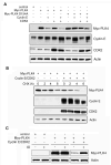
Figure 5. Cyclin E/CDK2 reduce PLK4 protein stability in a PLK4 kinase activity-dependent manner
(A) Immunoblot analysis of whole cell extracts from U-2 OS/centrin-GFP cells following transient transfection (48 h) with empty vector (control), Myc-tagged PLK4, Myc-tagged kinase-inactive mutant PLK4 D154A, cyclin E and/or CDK2. Note the decreased protein levels of wild-type PLK4 upon co-transfection with cyclin E or cyclin E/CDK2. No such reduction was detected in cells transfected with catalytically inactive mutant PLK4 D154A. Immunoblots for Actin are shown to demonstrate protein loading (A-C). (B) Immunoblot analysis of whole cell extracts from U-2 OS/centrin-GFP cells after transient transfection (48 h) with PLK4 alone or PLK4 and cyclin E/CDK2 and treatment with 30 μg/ml cycloheximide (CHX) for the indicated time intervals. Note the decreased PLK4 protein stability in the presence of ectopically expressed cyclin E/CDK2. (C) Immunoblot analysis of whole cell extracts from U-2 OS/centrin-GFP cells transiently transfected (24 h) with empty vector (control) or PLK4 alone or PLK4 in combination with cyclin E/CDK2 and treatment with 1 μM of the CDK inhibitor indirubin-3′-oxime for 24 h (IO). Note the stabilization of PLK4 protein in IO- treated cells in comparison to controls (0.1% DMSO).
Figure 6
Figure 6. CUL1 regulates PLK4 protein stability
(A) Immunofluorescence microscopic analysis of U-2 OS/centrin-GFP cells for endogenous PLK4 at 48 h after transfection of empty vector (control) or DN-CUL1 (left panels). Note centriole multiplication together with an excessive amount of endogenous PLK4 in DN-CUL1-transfected cells (bottom). Quantification of the fold-changes of PLK4 integrated signal intensities at maternal centrioles in U-2 OS/centrin-GFP cells transfected with empty vector or DN-CUL1 for 48 h or treated with 1 μM IO or 0.1% DMSO (control) for 48 h (right panel). (B) Immunoblot analysis of whole cell extracts from U-2 OS/centrin-GFP cells after transient transfection (48 h) with empty vectors (control) or PLK4 in combination with either cyclin E/CDK2 alone or cyclin E/CDK2 and DN-CUL1. Note the increase of PLK4 protein in the presence of DN-CUL1 in the last lane. (C) Immunoblot analysis of whole cell extracts from U-2 OS/centrin-GFP cells after transient transfection with empty vectors (control) or dominant-negative CUL1 (DN-CUL1) for 48 h followed by transfection with Myc-PLK4 for 24 h and treatment with 30 μg/ml cycloheximide (CHX) for the indicated time intervals. Note the increased protein stability of Myc-PLK4 in the presence of DN-CUL1 (6 h CHX, last lane). (D) Tentative model of centriole multiplication triggered by oncogenic stimuli such as c-MYC, E2F-1 or HPV-16 E7. These stimuli are known to deregulate cyclin E/CDK2 complexes and we show here that this leads to an aberrant recruitment of PLK4 to maternal centrioles. However, degradation of enzymatically active PLK4 (and low baseline protein expression) by CUL1-based SCF E3 ubiquitin ligase activity prevents the formation of supernumerary daughter centrioles. Only when PLK4 is overexpressed or its CUL1-mediated degradation is altered, centriole multiplication occurs. Hence, it is likely that oncogene-induced centriole multiplication, for example by HPV-16 E7, also involves impairment of CUL1-mediated protein degradation through mechanisms that remain to be determined. Genetic alterations such as deletion of the CUL1 locus in certain human malignancies are likewise to promote centriole duplication.

References

    1. Azimzadeh J, Bornens M. Structure and duplication of the centrosome. J Cell Sci. 2007;120(Pt 13):2139–42. - PubMed
    1. Strnad P, Gonczy P. Mechanisms of procentriole formation. Trends Cell Biol. 2008;18(8):389–96. - PubMed
    1. Nigg EA. Centrosome aberrations: cause or consequence of cancer progression? Nature Rev Cancer. 2002;2:1–11. - PubMed
    1. Duensing S, Munger K. Centrosome abnormalities, genomic instability and carcinogenic progression. Biochim Biophys Acta. 2001;2(8):M81–8. - VSports app下载 - PubMed
    1. Fukasawa K. Oncogenes and tumour suppressors take on centrosomes. Nat Rev Cancer. 2007;7(12):911–24. - PubMed

Publication types

MeSH terms

V体育2025版 - Substances

"VSports最新版本" LinkOut - more resources