Skip to main page content
U.S. flag

An official website of the United States government

Dot gov

The VSports app下载. gov means it’s official. Federal government websites often end in . gov or . mil. Before sharing sensitive information, make sure you’re on a federal government site. .

Https

The site is secure. The https:// ensures that you are connecting to the official website and that any information you provide is encrypted and transmitted securely V体育官网. .

. 2009 Jun 16;4(6):e5938.
doi: 10.1371/journal.pone.0005938.

"VSports app下载" Yersinia pestis endowed with increased cytotoxicity is avirulent in a bubonic plague model and induces rapid protection against pneumonic plague

Affiliations

Yersinia pestis endowed with increased cytotoxicity is avirulent in a bubonic plague model and induces rapid protection against pneumonic plague

Ayelet Zauberman et al. PLoS One. .

"V体育ios版" Abstract

An important virulence strategy evolved by bacterial pathogens to overcome host defenses is the modulation of host cell death. Previous observations have indicated that Yersinia pestis, the causative agent of plague disease, exhibits restricted capacity to induce cell death in macrophages due to ineffective translocation of the type III secretion effector YopJ, as opposed to the readily translocated YopP, the YopJ homologue of the enteropathogen Yersinia enterocolitica Oratio8. This led us to suggest that reduced cytotoxic potency may allow pathogen propagation within a shielded niche, leading to increased virulence. To test the relationship between cytotoxic potential and virulence, we replaced Y. pestis YopJ with YopP. The YopP-expressing Y. pestis strain exhibited high cytotoxic activity against macrophages in vitro. Following subcutaneous infection, this strain had reduced ability to colonize internal organs, was unable to induce septicemia and exhibited at least a 10(7)-fold reduction in virulence. Yet, upon intravenous or intranasal infection, it was still as virulent as the wild-type strain. The subcutaneous administration of the cytotoxic Y. pestis strain appears to activate a rapid and potent systemic, CTL-independent, immunoprotective response, allowing the organism to overcome simultaneous coinfection with 10,000 LD(50) of virulent Y. pestis. Moreover, three days after subcutaneous administration of this strain, animals were also protected against septicemic or primary pneumonic plague. Our findings indicate that an inverse relationship exists between the cytotoxic potential of Y. pestis and its virulence following subcutaneous infection. This appears to be associated with the ability of the engineered cytotoxic Y. pestis strain to induce very rapid, effective and long-lasting protection against bubonic and pneumonic plague. These observations have novel implications for the development of vaccines/therapies against Y. pestis and shed new light on the virulence strategies of Y. pestis in nature VSports手机版. .


VSports - Conflict of interest statement

Competing Interests: The authors have declared that no competing interests exist.

Figures

Figure 1
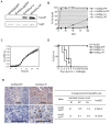
Figure 1. Expression of YopP in Y. pestis enhances its cytotoxicity in macrophages and reduces its virulence.
(A) Assessment of bacterial YopJ/P expression. Y. pestis recombinant strains were grown for 3 hrs at 37°C. Western blot analysis was conducted with anti-YopJ/P antibodies (upper lane) or anti-YopE antibodies (lower lane). Endogenous YopJ expression in both wild-type Kim53 and Kim53pGFP strains could be detected only under non-standard conditions of considerably longer exposure time (data not shown). (B) Cytotoxic effect of Y. pestis recombinant strains on J774A.1 macrophage cell line. Cells were infected with Kim53pGFP (circle), Kim53ΔJpGFP (X symbol), Kim53ΔJ+J (square) and Kim53ΔJ+P (triangle) at the indicated MOIs for 1 hour. Cell death was determined by LDH release test. (C) Growth curves of Kim53pGFP (circle), Kim53ΔJ+J (square) and Kim53ΔJ+P (triangle). Bacteria were inoculated to OD630 0.005 in heart infusion broth-containing microplate wells and incubated in a plate reader (Sunrise, Tecan) for an additional 22 hrs at 28°C. (D) Attenuation of YopP-expressing Y. pestis recombinant strain. Groups of mice were infected subcutaneously with 1×10² cfu of Kim53 (10 mice per group, white diamond), Kim53pGFP (5 mice per group, black circle) or Kim53ΔJ+J (5 mice per group, white square), or with 1×10⁶ cfu of Kim53ΔJ+P (5 mice per group, white triangle). Mortality rates were followed daily for 14 days after infection. (E) Spleen sections, isolated from mice 48 hrs after s.c. infection with 1×10⁴ cfu of Kim53pGFP or Kim53ΔJ+P, stained with anti-active caspase-3 antibodies (left panel). Scale bar = 50 µm. The arrow head indicates apoptotic cells. Caspase-positive cells in at least 20 random non-overlapping microscopic fields (magnification, ×1000) were counted (three mice per group, right panel).
Figure 2
Figure 2. Kim53ΔJ+P is deficient in dissemination to target organs and to the blood following subcutaneous infection.
Mice were infected subcutaneously with 1×10⁴ cfu of either Kim53pGFP (black symbols) or Kim53ΔJ+P (white symbols). Animals (4–5 per group) were sacrificed at 60 hours post-infection. Blood was collected, and the draining inguinal lymph nodes (ILN) and spleens were harvested, homogenized in 1 ml PBS and cultured on brain heart infusion agar plates at 28°C for 48 hrs. Values in Figure represent total bacterial loads in infected organs (cfu/organ), or bacterial concentration in blood (cfu/ml). LOD, limit of detection. Horizontal bars represent the average value of bacterial load in each case. Differences in bacterial concentrations in blood and internal organs were analyzed by the non-parametric Mann-Whitney test.
Figure 3
Figure 3. Kim53ΔJ+P is highly virulent following systemic or airway infection.
(A) Systemic infection (i.v. inoculation). Groups of 6 mice were infected intravenously with a dose of 2×10³ cfu of Kim53pGFP (circle) or Kim53ΔJ+P (triangle), and mortality was monitored daily for 14 days. (B) Airway infection (i.n. inoculation). Groups of 5 mice were infected intranasally with 6×10⁴ cfu of Kim53pGFP (circle) or Kim53ΔJ+P (triangle). (C) Dissemination of Y. pestis strains to blood and internal organs following intranasal infection. Groups of 5 mice were infected intranasally with 2×10⁵ cfu of Kim53pGFP (black symbol) or Kim53ΔJ+P (white symbol) and sacrificed 48 hours post-infection. Bacterial concentration in blood and total bacterial loads in lungs, mediastinal lymph nodes (MSLNs) and the spleen were determined as described in the legend to Figure 2.
Figure 4
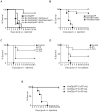
Figure 4. Subcutaneous administration of Kim53ΔJ+P protects mice coinfected with a virulent Y. pestis strain.
(A) Subcutaneous co-infections of the virulent Kim53pGFP and various Y. pestis strains were administered at a single site. Groups of 5 mice were infected at a single site located on the lower back with 1×10² cfu of Kim53pGFP alone (circle); a mixture of 1×10² cfu of Kim53pGFP with 1×10³ cfu of Kim53ΔJ+P (square); 1×10³ cfu of Kim53Δp10Δp70 (triangle); or 1×10³ cfu of Y. pestis pgm EV76 strain (diamond). Mortality was monitored daily for 14 days (B). Comparison of subcutaneous coinfection with Kim53ΔJ+P and Kim53pGFP administered at a single site (square) and at two separate sites (black triangle). Mice were injected in their lower back with a mixture of Kim53pGFP (1×10² cfu) and Kim53ΔJ+P (3×10³ cfu) for the single site. For the two separate sites, 10 mice were injected in the upper back with 3×10³ cfu of Kim53ΔJ+P and immediately thereafter with 1×10² cfu of Kim53pGFP in the lower back. Control mice were injected in the lower back with 1×10² cfu of Kim53pGFP (circle). (C) Subcutaneous infection of mice with Kim53ΔJ+P followed by intravenous challenge with virulent Y. pestis. In total, 8 mice were injected subcutaneously in the lower back with 104 cfu of Kim53ΔJ+P. Afterwards, 2×10³ cfu of the virulent Kim53pGFP were administered intravenously immediately (0 hr), or after 24 hrs or 72 hrs. Control mice were only infected intravenously with Kim53pGFP (circle). (D) Subcutaneous infection of mice with Kim53ΔJ+P followed by intranasal challenge with virulent Y. pestis. In total, 8 mice were injected subcutaneously with Kim53ΔJ+P as described in (C) and exposed intranasally to 6×10³ cfu (∼3LD50) of the virulent Kim53pGFP immediately (0 hr) or 72 hrs later. Mortality was monitored daily for 14 days. (E) Subcutaneous infections of KbDb−/− mice (class I MHC genes deleted) with 1×10³ cfu of Kim53pGFP (circle), 1×10³ cfu of Kim53ΔJ+P (open triangle) or 1×10⁴ cfu of Kim53ΔJ+P (closed triangle).
Figure 5
Figure 5. Y. pestis Kim53ΔJ+P strain confers prolonged protection against plague.
(A) Mice (7–8 per group) were infected subcutaneously with 1×10⁵ cfu of Kim53ΔJ+P (circle); EV76 (× symbol); or EV76ΔJ+P (triangle). Control animals were treated with saline (square). Animals were bled for ELISA determination of anti-F1 antibody titers 45 days following immunization. Titers following immunization with Kim53ΔJ+P ranged from 64,000 to 100,000, with GMT of 18,500, while titers for the EV76 or EV76ΔJ+P immunized animals ranged from 640 to less than 10, with GMT of 30 and 10, respectively. Animals were challenged subcutaneously with 1×10³ cfu (∼1×10³ LD50) of Y. pestis Kim53 (panel A) or intranasally with 8×10³ cfu (∼15 LD50) of Y. pestis Kim53 (panel B).

References

    1. Perry R, Fetherston J. Yersinia pestis-etiologic agent of plague. Clin Microbiol Rev. 1997;10:35–66. - PMC - PubMed
    1. Cornelis GR. Yersinia type III secretion: send in the effectors. The journal of Cell Biology. 2002;158:401–408. - "V体育官网" PMC - PubMed
    1. Marketon MM, DePaolo RW, DeBord KL, Jabri B, Schneewind O. Plague bacteria target immune cells during infection. Science. 2005;309:1739–1741. - PMC - PubMed
    1. Mota LJ, Cornelis GR. The bacterial injection kit: type III secretion systems. Ann Med. 2005;37:234–249. - PubMed
    1. Viboud GI, Bliska JB. Yersinia outer proteins: role in modulation of host cell signaling responses and pathogenesis. Annu Rev Microbiol. 2005;59:69–89. - PubMed

MeSH terms