Skip to main page content
U.S. flag

An official website of the United States government

Dot gov

The . gov means it’s official. Federal government websites often end in . gov or VSports app下载. mil. Before sharing sensitive information, make sure you’re on a federal government site. .

Https

The site is secure. The https:// ensures that you are connecting to the official website and that any information you provide is encrypted and transmitted securely V体育官网. .

. 2009 Jun 11;4(6):e5877.
doi: 10.1371/journal.pone.0005877.

"VSports最新版本" p63 promotes cell survival through fatty acid synthase

Affiliations

"V体育平台登录" p63 promotes cell survival through fatty acid synthase

Venkata Sabbisetti et al. PLoS One. .

Expression of concern in

Abstract

There is increasing evidence that p63, and specifically DeltaNp63, plays a central role in both development and tumorigenesis by promoting epithelial cell survival. However, few studies have addressed the molecular mechanisms through which such important function is exerted. Fatty acid synthase (FASN), a key enzyme that synthesizes long-chain fatty acids and is involved in both embryogenesis and cancer, has been recently proposed as a direct target of p53 family members, including p63 and p73. Here we show that knockdown of either total or DeltaN-specific p63 isoforms in squamous cell carcinoma (SCC9) or immortalized prostate epithelial (iPrEC) cells caused a decrease in cell viability by inducing apoptosis without affecting the cell cycle. p63 silencing significantly reduced both the expression and the activity of FASN. Importantly, stable overexpression of either FASN or myristoylated AKT (myr-AKT) was able to partially rescue cells from cell death induced by p63 silencing. FASN induced AKT phosphorylation and a significant reduction in cell viability was observed when FASN-overexpressing SCC9 cells were treated with an AKT inhibitor after p63 knockdown, indicating that AKT plays a major role in FASN-mediated survival. Activated AKT did not cause any alteration in the FASN protein levels but induced its activity, suggesting that the rescue from apoptosis documented in the p63-silenced cells expressing myr-AKT cells may be partially mediated by FASN VSports手机版. Finally, we demonstrated that p63 and FASN expression are positively associated in clinical squamous cell carcinoma samples as well as in the developing prostate. Taken together, our findings demonstrate that FASN is a functionally relevant target of p63 and is required for mediating its pro-survival effects. .


Conflict of interest statement

Competing Interests: The authors have declared that no competing interests exist.

Figures

Figure 1
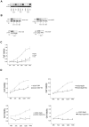
Figure 1. Silencing of either Total or ΔN specific p63 isoforms causes a decrease in cell viability.
A. Immunoblot analysis of p63 in SCC9 cells transfected with Tp63, Dp63 or Scr siRNAs. B. Immunoblot analysis of p63 in tetracycline-inducible clones of SCC9 (SCC9-Tp63, SCC9-Dp63), and iPrEC (iPrEC-Tp63 and iPrEC-Dp63) with or without treatment with tetracycline. C–D. Cell viability curves. Quantification of cell viability measured by MTT assay in SCC9 cells transfected with Tp63, Dp63 or Scr siRNAs (C), in SCC9-Tp63 and SCC9-Dp63 cells with or without treatment with tetracycline (D, upper panel) and in iPrEC-Tp63 and iPrEC-Dp63 cells with or without treatment with tetracycline (D, lower panel). Data are shown as means +/− standard error and are representative of three independent experiments.
Figure 2
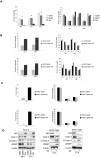
Figure 2. Silencing of either Total or ΔN specific p63 isoforms induces apoptosis with no significant changes in cell proliferation.
A–C. Flow cyometric analysis of SCC9 cells transfected with Tp63, Dp63 or Scr siRNAs (A), SCC9-Tp63 and SCC9-Dp63 cells with or without treatment tetracycline (B) and iPrEC-Tp63 and iPrEC-Dp63 cells with or without treatment tetracycline (C). Left panels show quantification (percentage) of the sub G1 population measured by PI staining. Right panels show the fractions of cells in the various phases of the cell cycle, as determined by BrdU incorporation. Data are shown as means +/− standard error and are representative of three independent experiments. D. Immunoblot analysis of apoptosis-related proteins in SCC9 cells transfected with Tp63, Dp63 or Scr siRNAs (left panel), in SCC9-Tp63 cells with or without treatment with tetracycline (middle panel) and in iPrEC-Tp63 cells with or without treatment with tetracycline (right panel).
Figure 3
Figure 3. p63 silencing induces a decrease in FASN expression and enzymatic activity.
A. Quantitation of FASN mRNA by real time RT-PCR in SCC9 cells transfected with Tp63 or Dp63 siRNA relative to cells transfected with Scr siRNA (left panel). Quantitation of FASN mRNA in SCC9-Tp63 and iPrEC-Tp63 cells treated with tetracycline relative to the correspondent untreated cells (middle and right panels, respectively). Data are shown as means +/− standard error and are representative of three independent experiments. B. Immunoblot analysis of FASN in SCC9 cells transfected with Tp63, Dp63 or Scr siRNAs (left panel), in SCC9-Tp63 cells with or without treatment with tetracycline (middle panel) and in iPrEC-Tp63 cells with or without treatment with tetracycline (right panel). C. Quantitation of C14 incorporation within cellular lipids in SCC9 cells transfected with Tp63 or Dp63 siRNA relative to cells transfected with Scr siRNA (left panel) and in SCC9-Tp63 and iPrEC-Tp63 cells treated with tetracycline relative to the correspondent untreated cells (middle and right panels, respectively). Data are shown as means +/− standard error and are representative of three independent experiments.
Figure 4
Figure 4. p63 silencing induces a decrease in AKT phosphorylation.
Immunoblot analysis of total AKT, phospho-AKT (Ser 473), and phospho-S6 (Ser 235/236) in SCC9 cells transfected with Tp63, Dp63 or Scr siRNAs (left panel), in SCC9-Tp63 cells with or without treatment with tetracycline (middle panel) and in iPrEC-Tp63 cells with or without treatment with tetracycline (right panel).
Figure 5
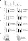
Figure 5. Increased FASN or AKT activity protects cells from apoptosis induced by p63 silencing.
A. Quantitation of apoptosis by flow cytometry using Annexin V assay (left panel) or PI staining (right panel) in EV-SCC9, mAKT-SCC9, and FASN-SCC9 cells transfected with either Tp63 or Scr siRNAs. B. Effects of p63 silencing on the viability of SCC9 and iPrEC cells overexpressing either FASN or empty vector. Upper panels: Quantification of cell viability in EV-SCC9 and FASN-SCC9 cells transfected with either Tp63 (left panel) or Dp63 (right panel) relative to cells transfected with Scr siRNA. Lower left panel: Quantification of cell viability in EV-SCC9 and FASN-SCC9 cells carrying inducible Tp63 shRNA (EV-SCC9-Tp63 and FASN-SCC9-Tp63) treated with tetracycline relative to untreated cells. Lower right panel: Quantification of cell viability in EV-iPrEC and FASN- iPrEC cells carrying inducible Tp63 shRNA (EV-iPrEC-Tp63 and FASN- iPrEC-Tp63) treated with tetracycline relative to untreated cells. C. Effects of p63 silencing on the viability of SCC9 and iPrEC cells overexpressing either Myr-AKT or empty vector. Upper panels: Quantification of cell viability in EV-SCC9 and mAKT-SCC9 cells transfected with either Tp63 (left panel) or Dp63 (right panel) relative to cells transfected with Scr siRNA. Lower left panel: Quantification of cell viability in EV-SCC9 and mAKT-SCC9 cells carrying inducible Tp63 shRNA (EV-SCC9-Tp63 and FASN-SCC9-Tp63) treated with tetracycline relative to untreated cells. Lower right panel: Quantification of cell viability in EV-iPrEC and mAKT-iPrEC cells carrying inducible Tp63 shRNA (EV-iPrEC-Tp63 and mAKT-iPrEC-Tp63) treated with tetracycline relative to untreated cells. Data are shown as means +/− standard error and are representative of three independent experiments. D. Immunoblot analysis of p63, FASN, phospho-AKT and c-PARP in EV-SCC9-Tp63 and FASN-SCC9-Tp63 cells with or without treatment with tetracycline. E. Inhibition of AKT signaling significantly affects FASN-mediated rescue of cell death induced by p63 silencing. The graph shows the viability of EV-SCC9-Tp63 with or without treatment with tetracycline and of FASN-SCC9-Tp63 and FASN-SCC9-Tp63 cells with or without treatment with an AKT inhibitor and/or tetracycline. Data are shown as means +/− standard error and are representative of three independent experiments.
Figure 6
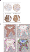
Figure 6. Co-localization of p63 and FASN in vivo.
A. Immunohistochemical staining for FASN and p63 in TMAs containing cores from HNSCC tissues. Two representative cases are shown: one case displays both low p63 and low FASN expression (upper panel) and one case displays both high p63 and high FASN expression (lower panel). B. Co-localization of p63 and FASN in the UGS epithelium and prostatic buds. (a) A coronal section of the UGS from a wild-type E18.5 embryo was double immunostained for p63 (Fast Red) and FASN (DAB) and counterstained with hematoxylin. Deconvolved images of the DAB (b) and Fast Red (c) color channels as well a pseudo-colorized image (c) are also shown. In the pseudo-colorized image, the DAB channel (FASN) is displayed in green, the Fast Red channel (p63) is displayed in red and the hematoxylin channel is displayed in blue. Expression of both FASN and p63 proteins is observed in the vast majority of the cells. The cells lining the UGS lumen are consistently negative for both FASN and p63.

References

    1. Yang A, Kaghad M, Wang Y, Gillett E, Fleming MD, et al. p63, a p53 homolog at 3q27-29, encodes multiple products with transactivating, death-inducing, and dominant-negative activities. Mol Cell. 1998;2:305–316. - PubMed
    1. Schmale H, Bamberger C. A novel protein with strong homology to the tumor suppressor p53. Oncogene. 1997;15:1363–1367. - "VSports在线直播" PubMed
    1. Osada M, Ohba M, Kawahara C, Ishioka C, Kanamaru R, et al. Cloning and functional analysis of human p51, which structurally and functionally resembles p53. Nat Med. 1998;4:839–843. - PubMed
    1. Yang A, Schweitzer R, Sun D, Kaghad M, Walker N, et al. p63 is essential for regenerative proliferation in limb, craniofacial and epithelial development. Nature. 1999;398:714–718. - V体育安卓版 - PubMed
    1. Signoretti S, Waltregny D, Dilks J, Isaac B, Lin D, et al. p63 is a prostate basal cell marker and is required for prostate development. Am J Pathol. 2000;157:1769–1775. - PMC - PubMed

Publication types

MeSH terms

LinkOut - more resources