V体育官网 - Skip to main page content
U.S. flag

An official website of the United States government

Dot gov

The . gov means it’s official VSports app下载. Federal government websites often end in . gov or . mil. Before sharing sensitive information, make sure you’re on a federal government site. .

Https

The site is secure V体育官网. The https:// ensures that you are connecting to the official website and that any information you provide is encrypted and transmitted securely. .

. 2009 Jun 15;69(12):4974-82.
doi: 10.1158/0008-5472.CAN-08-4671. Epub 2009 Jun 9.

ROC1/RBX1 E3 ubiquitin ligase silencing suppresses tumor cell growth via sequential induction of G2-M arrest, apoptosis, and senescence (V体育平台登录)

Affiliations

ROC1/RBX1 E3 ubiquitin ligase silencing suppresses tumor cell growth via sequential induction of G2-M arrest, apoptosis, and senescence

"V体育2025版" Lijun Jia et al. Cancer Res. .

Abstract

Regulator of Cullins-1 (ROC1) or Ring Box Protein-1 (RBX1) is a RING component of SCF (Skp-1, cullins, F-box proteins) E3 ubiquitin ligases, which regulate diverse cellular processes by targeting a variety of substrates for degradation. However, little is known about the role of ROC1 in human cancer. Here, we report that ROC1 is ubiquitously overexpressed in primary human tumor tissues and human cancer cell lines. ROC1 silencing by siRNA significantly inhibited the growth of multiple human cancer cell lines via induction of senescence and apoptosis as well as G(2)-M arrest. Senescence induction is coupled with DNA damage in p53/p21- and p16/pRB-independent manners. Apoptosis is associated with accumulation of Puma and reduction of Bcl-2, Mcl-1, and survivin; and G(2)-M arrest is associated with accumulation of 14-3-3sigma and elimination of cyclin B1 and Cdc2. In U87 glioblastoma cells, these phenotypic changes occur sequentially upon ROC1 silencing, starting with G(2)-M arrest, followed by apoptosis and senescence VSports手机版. Thus, ROC1 silencing triggers multiple death and growth arrest pathways to effectively suppress tumor cell growth, suggesting that ROC1 may serve as a potential anticancer target. .


Figures

Figure 1
Figure 1. The expression of ROC1 in human tumors and normal counterparts
Tumor tissue arrays containing multiple normal and tumor tissues from different organs were stained with a specific ROC1 antibody on the DAKO AutoStainer using the DakoCytomation EnVision+ System-HRP (DAB) detection kit, and counterstained with hematoxylin (Surgipath). Shown are representative images of normal vs. tumor tissues of lung, liver, and breast with two magnifications (A). Lung tumor tissue arrays containing normal lung and tumor tissues were stained for ROC1 expression. Stained normal and tumor tissues were classified into five groups according to staining intensity of each tissue and samples with different staining intensity were grouped and tabulated (B&C).
Figure 2
Figure 2. ROC1 silencing inhibited the growth of human cancer cells
U87 and H1299 cells were infected with LT-CONT and LT-ROC1 for 72 hrs and cells were then split for following assays: ROC1 silencing by immunoblotting (A&B, top), cell proliferation (A&B, middle). clonogenic survival (A&B, bottom), and soft agar anchorage-independent growth in H1299 cells (C). Shown is mean ± SEM from three independent experiments, each run in quadruplicate (A&B, middle) or triplicate (A&B, bottom and C).
Figure 3
Figure 3. ROC1 silencing induced cell senescence in p53/p21- and p16/pRB-independent manner
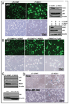
U87 cells were infected with LT-ROC1, along with LT-CONT control for 120 hrs, followed by morphological observation under green fluorescence (A, top left), SA–β–gal staining (A, bottom left) and IB analysis (A, top right). U87 cells were infected with LT-CONT/LT-CONT, LT-CONT/LT-ROC1 or LT-ROC1/LT-p53 for 120 hrs, followed by IB analysis (A, bottom right), morphological observation under green fluorescence (B, top), SA–β–gal staining (B, bottom). p16 and pRB expression in ROC1-silenced U87 cells (C, top). pRB expression in HeLa and MDA-MB-468 cells (C, bottom). HeLa and MDA-MB-468 cells were infected with LT-ROC1 along with LT-CONT for 72 hrs, cells were split and cultured for 120 hrs, followed by SA–β–gal staining (D). Representative results of three independent experiments are shown.
Figure 4
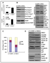
Figure 4. ROC1 silencing induced apoptosis and G2/M arrest in U87 cells
U87 and H1299 cells were infected with LT-ROC1, along with LT-CONT control for 120 hrs (U87), or for 72 hrs, then split and cultured for 72 hrs (H1299), followed by FACS analysis. Shown is mean value ± SEM from three independent experiments (A&C). U87 cells after infection for 120 hrs were subjected to immunoblotting analysis for indicated proteins (B&D). Representative results of three independent experiments were shown.
Figure 5
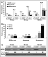
Figure 5. ROC1 silencing sequentially induced G2/M arrest, apoptosis and senescence
U87 cells were infected with LT-ROC1 or LT-CONT. Cells were harvested at 72, 96, and 120 hrs post infection. One portion was used for FACS analysis to measure the % of cells arrested at the G2/M or undergoing apoptosis (Sub-G1 fraction). Second portion was used for immunoblotting analysis for the degree of ROC1 silencing. The third set of cells in cover slid was subject to SA–β–gal staining for senescence. (A) Percentage of cells arrested at the G2/M phase, or undergoing apoptosis or senescence. (B) Fold-increase in each category of ROC1-silenced cells over the control cells. (C) The levels of ROC1 silencing at each time point measured by immunoblotting analysis.
Figure 6
Figure 6. ROC1 silencing induced DNA damage
U87 cells were infected with LT-ROC1 or LT-CONT for 120 hrs with the treatment by DMSO or 25 µM etoposide at the last 12 hrs of culture. Cells were either stained with anti-phosphor-γH2AX Ab or DAPI for cellular nuclear (A), or subjected to IB analysis (B). U87 cells were infected with LT-ROC1 or LT-CONT for 48 hrs, 72 hrs, 96 hrs or 120 and subjected to IB analysis (C). The relative levels of phosphor-γH2AX were quantified by densitometry analysis using Image J1.410 image processing software (bottom panel).

References

    1. Skowyra D, Craig KL, Tyers M, Elledge SJ, Harper JW. F-box proteins are receptors that recruit phosphorylated substrates to the SCF ubiquitin-ligase complex [see comments] Cell. 1997;91:209–219. - PubMed
    1. Ohta T, Michel JJ, Schottelius AJ, Xiong Y. ROC1, a homolog of APC11, represents a family of cullin partners with an associated ubiquitin ligase activity. Mol Cell. 1999;3:535–541. - PubMed
    1. Kamura T, Conrad MN, Yan Q, Conaway RC, Conaway JW. The Rbx1 subunit of SCF and VHL E3 ubiquitin ligase activates Rub1 modification of cullins Cdc53 and Cul2. Genes Dev. 1999;13:2928–2933. - PMC - PubMed
    1. Tan P, Fuchs SY, Chen A, et al. Recruitment of a ROC1-CUL1 ubiquitin ligase by Skp1 and HOS to catalyze the ubiquitination of IkBa. Mol Cell. 1999;3:527–533. - PubMed
    1. Seol JH, Feldman RMR, Zachariae WZ, et al. Cdc53/cullin and the essential Hrt1 RING-H2 subunit of SCF define a ubiquitin ligase module that activates the E2 enzyme Cdc34. Genes & Dev. 1999;13:1614–1626. - PMC - PubMed

Publication types

"VSports在线直播" LinkOut - more resources