Skip to main page content
U.S. flag

An official website of the United States government

Dot gov

The . gov means it’s official. Federal government websites often end in . gov or . mil. Before sharing sensitive information, make sure you’re on a federal government site. VSports app下载.

Https

The site is secure. The https:// ensures that you are connecting to the official website and that any information you provide is encrypted and transmitted securely V体育官网. .

. 2009 Oct;13(10):4205-18.
doi: 10.1111/j.1582-4934.2008.00539.x.

Combination of taxol and Bcl-2 siRNA induces apoptosis in human glioblastoma cells and inhibits invasion, angiogenesis and tumour growth

Affiliations

Combination of taxol and Bcl-2 siRNA induces apoptosis in human glioblastoma cells and inhibits invasion, angiogenesis and tumour growth (V体育2025版)

Joseph George et al. J Cell Mol Med. 2009 Oct.

Abstract

Taxol is a powerful chemotherapeutic agent that binds to microtubules to prevent tumour cell division. However, a traditional high dose of taxol may also induce apoptosis in normal cells. The anti-apoptotic molecule Bcl-2 is up-regulated in tumour cells to prevent apoptosis. We designed this study to determine whether use of a low dose of taxol and anti-apoptotic Bcl-2 gene silencing would effectively induce apoptosis in human glioblastoma U251MG cells and also inhibit invasion, angiogenesis and intracranial as well as subcutaneous tumour growth. We treated the cells with either 100 nM taxol or transfected with a plasmid vector expressing Bcl-2 siRNA or both agents together for 72 h. Knockdown of Bcl-2 potentiated efficacy of taxol for cell death. Fluorescence-activated cell sorting analysis, double immunofluorescent staining and TUNEL assay demonstrated apoptosis in about 70% of the cells after treatment with the combination of taxol and Bcl-2 siRNA. In vitro Matrigel invasion assay demonstrated dramatic decrease in glioblastoma cell invasion and in vivo angiogenesis assay showed complete inhibition of neovascularization in athymic nude mice after treatment with the combination. Further, treatment with the combination of taxol and Bcl-2 siRNA caused suppression of intracranial tumour growth and subcutaneous solid tumour development. In conclusion, our results indicate that the combination of taxol and Bcl-2 siRNA effectively induces apoptosis and inhibits glioblastoma cell invasion, angiogenesis and intracranial as well as subcutaneous tumour growth. Therefore, the combination of a low dose of taxol and Bcl-2 siRNA is a promising therapeutic strategy for controlling the aggressive growth of human glioblastoma VSports手机版. .


Figures
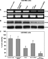
Fig 1
Fig 1
Expression of Bcl-2 at mRNA and protein levels in U251MG cells. Treatment (72 hrs) of cells: control, 100 nM taxol, transfection with a plasmid vector carrying the scrambled siRNA cDNA, transfection with a plasmid vector carrying the Bcl-2 siRNA cDNA and taxol + Bcl-2 siRNA. In determination of both RNA and protein levels, GAPDH was used as an internal control. (A) Semi-quantitative RT-PCR for examination of Bcl-2 mRNA. (B) Western blotting for examination of Bcl-2 protein. (C) MTT assay for determination of percent changes in cell viability. For MTT assay, the cells were treated for 48 hrs. Data are representative of six independent experiments in duplicate (*P < 0.001 when compared with the control mean values and #P < 0.001 when compared with taxol or Bcl-2 siRNA mean values).
Fig 2
Fig 2
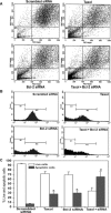
FACS analysis for detection of apoptotic cells and DNA fragmentation. Treatment (72 hrs) of cells: transfection with a plasmid vector expressing scrambled siRNA (treated control), 100 nM taxol, transfection with a plasmid vector expressing Bcl-2 siRNA and taxol + Bcl-2 siRNA. (A) FACS dot plots of U251MG cells. Before FACS analysis, the cells were treated with 50 μg/ml propidium iodide for 30 min. at 4°C in dark. Population in the M1 area represents the apoptotic/dead cells. (B) FACS histograms of U251MG cells. The prominent increase in population of cells in the sub-G1 phase (M1) indicated increase in apoptosis after treatment with taxol or Bcl-2 siRNA or both. (C) Quantitative presentation of DNA fragmentation data from FACS analysis to indicate percent changes in live and apoptotic cells. Data are representative of four independent experiments (*P < 0.001 when compared with scrambled siRNA treatment mean values and #P < 0.001 when compared with taxol or Bcl-2 siRNA treatment mean values).
Fig 3
Fig 3
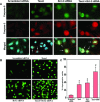
In situ stainings for biochemical markers of apoptosis in U251MG cells. Treatment (72 hrs) of cells: transfection with a plasmid vector expressing scrambled siRNA (treated control), 100 nM taxol, transfection with a plasmid vector expressing Bcl-2 siRNA and taxol + Bcl-2 siRNA. (A) Double immunofluorescent stainings to examine active fragments of caspase-9 and caspase-3. The cells were incubated overnight at 4°C with specific antibodies for active fragments of caspase-9 and caspase-3, washed and then treated with FITC conjugated and Texas red conjugated secondary antibodies at room temperature for 1 hr. Hoechst 33342 was used to counterstain the nucleus. Merged microphotographs demonstrated simultaneous expression of active fragments of caspase-9 and caspase-3 as well as disintegration of nucleus in the apoptotic cells (shown with arrows). (B) Fluorescent TUNEL assay for detection of apoptotic cells. Treatment with combination of taxol and Bcl-2 siRNA resulted in more apoptotic cell death than either treatment alone. (C) Quantitation of TUNEL-positive cells. Data are representative of four independent experiments (*P < 0.001 when compared with the scrambled siRNA treatment mean values and #P < 0.001 when compared with taxol or Bcl-2 siRNA treatment mean values).
Fig 4
Fig 4
In vitro Matrigel invasion assay using U138MG and U251MG cells. Treatment (72 hrs) of cells: transfection with a plasmid vector expressing scrambled siRNA (treated control), 100 nM taxol, transfection with a plasmid vector expressing Bcl-2 siRNA and taxol + Bcl-2 siRNA. Invasion assays were carried out in 12-well transwell inserts of polycarbonate filters. After 48 hrs incubation at 37°C in a CO2 incubator, the membranes were collected and stained with HEMA. The number of cells that migrated to the undersurface of the membrane were examined under a microscope, counted and photographed. (A) The changes in capability of invasion of U138MG cells after the treatments. (B) Quantitative evaluation of invading U138MG cells. (C) The changes in capability of invasion of U251MG cells after the treatments. (D) Quantitative evaluation of invading U251MG cells. The quantitative data are presented as mean ± S.D. of cells from 10 randomly selected microscopic fields from three independent wells (*P < 0.001 when compared with the scrambled siRNA treatment mean values and #P < 0.001 when compared with taxol or Bcl-2 siRNA treatment mean values).
Fig 5
Fig 5
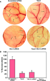
In vivo angiogenesis (dorsal skinfold) assay. Treatment (72 hrs) of cells: transfection with a plasmid vector expressing scrambled siRNA (treated control), 100 nM taxol, transfection with a plasmid vector expressing Bcl-2 siRNA and taxol + Bcl-2 siRNA. The U251MG cells (2 × 10⁵) from each treatment were suspended in 200 μl of serum-free medium and injected into a diffusion chamber. Then, diffusion chambers were surgically implanted under the dorsal skin of nude mice and left for 10 days. (A) In vivo angiogenesis in terms of development of neovasculature. Strong development of tumour-induced neovasculature (TN) with curved thin structures (as indicated by TN arrows) arising from pre-existing vasculature (PV) with relatively straight structures (as indicated by PV arrows) was observed in nude mice with U251MG cells untreated (not shown) and transfected with scrambled siRNA vector. The formation of such neovasculature was considerably reduced in nude mice with U251MG cells treated with taxol or Bcl-2 siRNA and completely inhibited in nude mice with U251MG cells treated with combination of taxol and Bcl-2 siRNA. (B) Quantitative presentation of neovasculature to indicate the extent of in vivo angiogenesis. The measurement of the TN was performed with the help of an ocular micrometer. Values are presented as mean ± S.D. of TN in six animals from each treatment group (*P <0.001 when compared with the scrambled siRNA treatment mean values).
Fig 6
Fig 6
Longitudinal bioluminescence imaging of intracranial tumour growth in nude mice. The U251MG cells (1 × 10⁶) stably transfected with luciferase gene were used for treatments and implantations in nude mice. Treatment (72 hrs) of U251MG cells: control (untreated control), transfection with a plasmid vector expressing scrambled siRNA (treated control), 100 nM taxol, transfection with a plasmid vector expressing Bcl-2 siRNA and taxol + Bcl-2 siRNA. The cells from each treatment were harvested and 1 × 10⁶ cells were suspended in 10 μl of serum-free medium and injected into the cerebrum of nude mice with the help of a stereotaxic apparatus. On day 3 onwards, the mice were injected intraperitoneally with nothing (control) or scrambled siRNA vector (treated control) or taxol or Bcl-2 siRNA vector or taxol + Bcl-2 siRNA vector for 20 days on alternate days. On days 7, 14 and 21, the mice were injected with luciferin for visualization of bioluminescence from luciferase activity to detect the extent of intracranial tumour growth in nude mice. The data are representative of six mice in each treatment group.
Fig 7
Fig 7
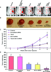
Examination of subcutaneous tumourigenesis and solid tumour development in nude mice. Treatment (72 hrs) of U251MG cells: control (untreated control), transfection with a plasmid vector expressing scrambled siRNA (treated control), 100 nM taxol, transfection with a plasmid vector expressing Bcl-2 siRNA and taxol + Bcl-2 siRNA. (A) Inhibition of subcutaneous tumourigenesis in nude mice. The U251MG cells (carrying the luciferase gene) were treated as mentioned above and the, injected under the dorsal of skin of nude mice. Beginning from day 3, the mice received intraperitoneal injection of nothing (control) or scrambled siRNA vector or taxol or Bcl-2 siRNA vector or taxol + Bcl-2 siRNA vector on alternate days for 20 days. On day 21, the mice were injected with luciferin and visualized for luciferase activity. The data are representative of six mice in each treatment group. (B) Inhibition of solid tumour development in the subcutaneous tissue of nude mice. The U251MG cells (not carrying the luciferase gene) were treated as mentioned above, harvested and suspended in an equal volume of the Matrigel, and 100 μl of this suspension (5 × 10⁶ cells) was injected subcutaneously in nude mice. The animals were left for 2 weeks without any treatment. Afterwards, the mice received intraperitoneal injection of nothing (control) or scrambled siRNA vector or taxol or Bcl-2 siRNA vector or taxol + Bcl-2 siRNA vector on alternate days for 4 weeks. At the end of the sixth week, the tumours were surgically removed, weighed and photographed. (C) Longitudinal measurement of tumour volume in nude mice using a digital vernier caliper. The data are presented as mean ± S.D. of six animals in each treatment group. (D) Measurement of tumour weight following the treatments. The data are presented as mean ± S.D. of six animals in each treatment group (*P < 0.001 when compared with the scrambled siRNA treatment mean values and #P < 0.001 when compared with taxol or Bcl-2 siRNA treatment mean values).

References (VSports在线直播)

    1. Pulkkanen KJ, Yla-Herttuala S. Gene therapy for malignant glioma: current clinical status. Mol Ther. 2005;12:585–98. - PubMed
    1. Donaldson SS, Laningham F, Fisher PG. Advances toward an understanding of brainstem gliomas. J Clin Oncol. 2006;24:1266–72. - PubMed
    1. Hood JD, Cheresh DA. Role of integrins in cell invasion and migration. Nat Rev Cancer. 2002;2:91–100. - PubMed (VSports最新版本)
    1. Zagzag D, Friedlander DR, Margolis B, et al. Molecular events implicated in brain tumor angiogenesis and invasion. Pediatr Neurosurg. 2000;33:49–55. - PubMed
    1. Kitada S, Pedersen IM, Schimmer AD, et al. Dysregulation of apoptosis genes in hematopoietic malignancies. Oncogene. 2002;21:3459–74. - PubMed

Publication types

MeSH terms

"V体育平台登录" LinkOut - more resources