Skip to main page content
U.S. flag

An official website of the United States government

Dot gov

The . gov means it’s official. Federal government websites often end in VSports app下载. gov or . mil. Before sharing sensitive information, make sure you’re on a federal government site. .

Https

The site is secure V体育官网. The https:// ensures that you are connecting to the official website and that any information you provide is encrypted and transmitted securely. .

. 2009 Aug 1;47(3):312-20.
doi: 10.1016/j.freeradbiomed.2009.05.012. Epub 2009 May 15.

VSports手机版 - Glutathione peroxidase 4 differentially regulates the release of apoptogenic proteins from mitochondria

Affiliations

Glutathione peroxidase 4 differentially regulates the release of apoptogenic proteins from mitochondria

Hanyu Liang et al. Free Radic Biol Med. .

Abstract

Glutathione peroxidase 4 (Gpx4) is a unique antioxidant enzyme that repairs oxidative damage to biomembranes. In this study, we examined the effects of Gpx4 on the release of various apoptogenic proteins from mitochondria using transgenic mice overexpressing Gpx4 [Tg(GPX4(+/0))] and mice deficient in Gpx4 (Gpx4+/- mice) VSports手机版. Diquat exposure triggered apoptosis that occurred through an intrinsic pathway and resulted in the mitochondrial release of cytochrome c (Cyt c), Smac/DIABLO, and Omi/HtrA2 in the liver of wild-type (Wt) mice. Liver apoptosis and Cyt c release were suppressed in Tg(GPX4(+/0)) mice but exacerbated in Gpx4+/- mice; however, neither the Tg(GPX4(+/0)) nor the Gpx4+/- mice showed any alterations in the levels of Smac/DIABLO or Omi/HtrA2 released from mitochondria. Submitochondrial fractionation data showed that Smac/DIABLO and Omi/HtrA2 existed primarily in the intermembrane space and matrix, whereas Cyt c and Gpx4 were both associated with the inner membrane. In addition, diquat exposure induced cardiolipin peroxidation in the liver of Wt mice; the levels of cardiolipin peroxidation were reduced in Tg(GPX4(+/0)) mice but elevated in Gpx4+/- mice. These data suggest that Gpx4 differentially regulates apoptogenic protein release owing to its inner membrane location in mitochondria and its ability to repair cardiolipin peroxidation. .


Figures

Figure 1
Figure 1. The effect of Gpx4 on diquat induced apoptosis
Mice were treated with diquat (50 mg/kg). The levels of liver apoptosis and caspase 9 activation were determined as described in the Experimental Procedures. Panel A: Time course for the induction of apoptosis in the livers of Wt mice. The asterisks show those values that are significantly different from untreated controls. Panel B: Level of apoptosis in the livers of Wt (open bars) and Tg(GPX4+/0) (solid bars) mice untreated or treated with diquat for 6 hours. Panel C: Level of apoptosis in the livers of Wt (hatched bars) and Gpx4+/− mice (shaded bars) mice untreated or treated with diquat for 6 hours. Panel D: Photograph of a representative western blot showing cleaved caspase-9 (p17) in the liver homogenates isolated from Wt and Tg(GPX4+/0) mice untreated or treated with diquat for 6 hours. Panel E: Quantification of cleaved caspase-9 (p17) from Wt (open bars) and Tg(GPX4+/0) (solid bars) mice as determined from the Western blots. All values are the mean ± SEM of data obtained from 3–4 mice. The asterisks in Panels B, C, and E show those values that are significantly different.
Figure 2
Figure 2. The levels of Gpx4 in Tg(GPX4+/0) and Gpx4+/− mice
Tg(GPX4+/0) and Gpx4+/− mice and their corresponding Wt littermates were either untreated or treated with diquat. Gpx4 protein levels in the liver were determined by Western blotting as described in the Experimental Procedures. Panel A: Quantification of Gpx4 protein levels in the liver of Tg(GPX4+/0) mice (solid bars) and their Wt littermates (open bars) as determined from the Western blotting. Panel B: Quantification of Gpx4 protein levels the liver of Gpx4+/− mice (shaded bars) and their Wt littermates (hatched bars) as determined from the Western blotting. Values are mean ± SEM of data obtained from 3–4 mice. The asterisks indicate those differences that are significantly different.
Figure 3
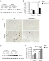
Figure 3. Effect of Gpx4 on diquat-induced cyt. c release
Tg(GPX4+/0) and Gpx4+/− mice and their corresponding Wt littermates were either untreated or treated with diquat. Cyt. c release was determined by Western blotting and immunohistochemistry as described in the Experimental Procedures. Panel A: Photograph of a representative Western blotting showing cyt. c release into the cytosol in Tg(GPX4+/0) mice and their Wt littermates. Panel B: Quantification of cyt.c release in Tg(GPX4+/0) mice (solid bars) and their Wt littermates (open bars) as determined from the Western blotting. Panel C: Photograph of representative liver sections from Wt and Tg(GPX4+/0) mice. Sections were immunostained with the anti-cyt. c antibody. Arrows show examples of cyt. c positive cells. Original magnification: ×200. Scale bar=50 µm. Panel D: Photograph of a representative Western blotting showing cyt. c release into the cytosol in Gpx4+/− mice and their Wt littermates. Panel E: Quantification of cyt.c release in Gpx4+/− mice (shaded bars) and their Wt littermates (hatched bars) as determined from the Western blotting. Values are mean ± SEM of data obtained from 3–4 mice. The asterisks indicate those differences that are significantly different.
Figure 4
Figure 4. Effect of Gpx4 on diquat-induced Smac/DIABLO release from liver mitochondria
Tg(GPX4+/0) and Gpx4+/− mice and their corresponding Wt littermates were either untreated or treated with diquat. Smac/DIABLO release was determined by measuring the levels of Smac/DIABLO in the cytosol using Western blotting as described in the Experimental Procedures. Panel A: Photograph of a representative Western blot showing Smac/DIABLO release into the cytosol from the Tg(GPX4+/0) mice and their Wt littermates. Panel B: Quantification of Smac/DIABLO release in the Tg(GPX4+/0) mice (solid bars) and their Wt littermates (open bars) as determined from the Western blotting. Panel C: Photograph of a representative Western blotting showing Smac/DIABLO release into the cytosol from the Gpx4+/− mice and their Wt littermates. Panel D: Quantification of Smac/DIABLO release in the Gpx4+/− mice (shaded bars) and their Wt littermates (hatched bars) as determined from the Western blotting. All values are mean ± SEM of data obtained from 3–4 mice. The asterisks indicate the differences that are significantly different.
Figure 5
Figure 5. Effect of Gpx4 on diquat-induced Omi/HtrA2 release from liver mitochondria
Tg(GPX4+/0) and Gpx4+/− mice and their corresponding Wt littermates were either untreated or treated with diquat. Omi/HtrA2 release was determined by measuring the levels of Omi/HtrA2 in the cytosol using Western blotting as described in the Experimental Procedures. Panel A: Photograph of a representative Western blot showing Omi/HtrA2 release into the cytosol from the Tg(GPX4+/0) mice and their Wt littermates. Panel B: Quantification of Omi/HtrA2 release in the Tg(GPX4+/0) mice (solid bars) and their Wt littermates (open bars) as determined from the Western blotting. Panel C: Photograph of a representative Western blotting showing Omi/HtrA2 release into the cytosol from the Gpx4+/− mice and their Wt littermates. Panel D: Quantification of Omi/HtrA2 release in the Gpx4+/− mice (shaded bars) and their Wt littermates (hatched bars) as determined from the Western blotting. All values are mean ± SEM of data obtained from 3–4 mice. The asterisks indicate the differences that are significantly different.
Figure 6
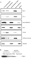
Figure 6. Submitochondrial localization of Gpx4 and apoptogenic proteins
Submitochondrial fractions were obtained as described in the Experimental Procedures. Localizations of Gpx4 and apoptogenic proteins are shown by Western blotting. Panel A: Photograph of a representative Western blot showing the localization of Gpx4 and apoptogenic proteins in the mitochondria. Panel B: Photograph of a representative Western blotting showing the integral membrane association of Gpx4 with the inner membrane.
Figure 7
Figure 7. Effect of Gpx4 on CL peroxidation
Tg(GPX4+/0) and Gpx4+/− mice and their corresponding Wt littermates were either untreated or treated with diquat and CL peroxidation determined by measuring mitochondrial bound NAO as described in the Experimental Procedures. Panel A: Quantification of CL peroxidation in Tg(GPX4+/0) mice (solid bars) and their Wt littermates (open bars). Panel B: Quantification of CL peroxidation in Gpx4+/− mice (shaded bars) and their Wt littermates (hatched bars). All values are mean ± SEM of data obtained from 4 mice. The asterisks indicate the differences that are significantly different.

References

    1. Reed JC. Mechanisms of apoptosis. Am. J. Pathol. 2000;157:1415–1430. - PMC - PubMed
    1. Green DR. Apoptotic pathways: the roads to ruin. Cell. 1998;94:695–698. - PubMed (VSports app下载)
    1. Spierings D, McStay G, Saleh M, Bender C, Chipuk J, Maurer U, Green DR. Connected to death: the (unexpurgated) mitochondrial pathway of apoptosis. Science. 2005;310:66–67. - PubMed
    1. Jabs T. Reactive oxygen intermediates as mediators of programmed cell death in plants and animals. Biochem. Pharmacol. 1999;57:231–245. - PubMed
    1. Skulachev VP. Cytochrome c in the apoptotic and antioxidant cascades. FEBS Lett. 1998;423:275–280. - PubMed

Publication types

MeSH terms

LinkOut - more resources