<u lang="GrYN6SS1"></u><center lang="xySXLoVm"></center> Skip to main page content
U.S. flag

An official website of the United States government

Dot gov

The . gov means it’s official. Federal government websites often end in . gov or . mil VSports app下载. Before sharing sensitive information, make sure you’re on a federal government site. .

Https

The site is secure. The https:// ensures that you are connecting to the official website and that any information you provide is encrypted and transmitted securely V体育官网. .

. 2009 May 15;182(10):5938-48.
doi: 10.4049/jimmunol.0802212.

"VSports在线直播" Central memory CD8+ T cells induce graft-versus-host disease and mediate graft-versus-leukemia

Affiliations

Central memory CD8+ T cells induce graft-versus-host disease and mediate graft-versus-leukemia

Hong Zheng et al. J Immunol. .

Abstract

In allogeneic hemopoietic stem cell transplantation, mature donor alphabeta T cells in the allograft promote T cell reconstitution in the recipient and mediate the graft-vs-leukemia (GVL) effect. Unfortunately, donor T cells can attack nonmalignant host tissues and cause graft-vs-host disease (GVHD). It has previously been shown that effector memory T cells not primed to alloantigen do not cause GVHD yet transfer functional T cell memory and mediate GVL. Recently, central memory T cells (T(CM)) have also been reported to not cause GVHD. In contrast, in this study, we demonstrate that purified CD8(+) T(CM) not specifically primed to alloantigens mediate GVHD in the MHC-mismatched C57BL/6 (B6)-->BALB/c and the MHC-matched, multiple minor histocompatibility Ag-mismatched C3H. SW-->B6 strain pairings. CD8(+) T(CM) and naive T cells (T(N)) caused similar histological disease in liver, skin, and bowel VSports手机版. B6 CD8(+) T(CM) and T(N) similarly expanded in BALB/c recipients, and the majority of their progeny produced IFN-gamma upon restimulation. However, in both models, CD8(+) T(CM) induced milder clinical GVHD than did CD8(+) T(N). Nonetheless, CD8(+) T(CM) and T(N) were similarly potent mediators of GVL against a mouse model of chronic-phase chronic myelogenous leukemia. Thus, in contrast to what was previously thought, CD8(+) T(CM) are capable of inducing GVHD and are substantially different from T(EM) but only subtly so from T(N). .


Conflict of interest statement

Disclosures

The authors have no conflicting financial interests.

Figures (V体育官网)

Figure 1.
Figure 1.. CD8+ TCM induce milder but definite GVHD in the B6→BALB/c strain pairing.
A. Cell sorting. TN and TCM CD8 cells were purified and sorted from splenocytes as described in Methods. We gated on CD8+ cells (first panel), which were sorted into TN and TCM based on the expression of CD62L and CD44. B and C. GVHD in B6→BALB/c model (data combined from 2 experiments). BALB/c mice were lethally irradiated and reconstituted with TCD B6 BM, with no T cells or 1×10⁶ CD8+ TN or TCM. B. Weight change. P<0.005 at days 5, 8, 12 and 14 comparing recipients of BM versus CD8+ TN or TCM; P=0.013 comparing BM alone versus TN on day 20. P<0.01 at day 20, 23, 27, 30, 34, 37 and 40 comparing recipients of CD8+ TN versus CD8+ TCM. C. Liver pathology. Each symbol represents a score from an individual mouse; horizontal lines are mean scores. P=0.0018 comparing BM and CD8+ TN recipients; P=0.0085 comparing BM and CD8+ TCM recipients. P=0.086 comparing CD8+ TN versus CD8+ TCM. D and E. BALB/c mice were irradiated and reconstituted with TCD B6 BM, with no T cells, 1×10⁶ TN, 1×10⁶ TCM or 2×10⁴ TN (TN control) B6 CD8 cells. D. Weight change. E. Liver pathology scores. P<0.005 for recipients of TN or TCM versus TN control. P value is not significant between BM and TN control recipients.
Figure 2.
Figure 2.. CD8+ TCM induce milder but definite GVHD in C3H.SW→B6 strain pairing.
Data combined from 3 experiments. B6 mice were lethally irradiated and reconstituted with TCD C3H.SW BM, with no T cells, 1.5×10⁶ CD8+ TN or TCM. A. Survival. P=0.0029 comparing BM and TN; P=0.2138 comparing BM and TCM; P=0.054 comparing TN and TCM. B. Weight change. P<0.001 at days 26, 30, 34, 37 and 43 comparing recipients of BM only versus CD8+ TN; P<0.05 at days 26 and 43 comparing recipients of TN versus TCM. P values not significant at any time-point comparing BM alone versus TCM. C. Pathology scores. Each symbol represents a score from an individual mouse; horizontal lines are mean scores. P values are noted below each panel.
Figure 3.
Figure 3.. CD8+ TN and TCM infiltrate small bowel in the C3H.SW→B6 strain pairing.
Frozen sections of small bowel from recipients of only TCD BM and TN or TCM recipients with confirmed histologic GVHD (by blinded scoring) were stained with anti-CD8 (Alexa647; rendered in green). DAPI-stained nuclei are rendered in blue. Shown in A are representative sections from two recipients of TN, TCM or only TCD BM. In TN and TCM recipients, CD8 cells percolate through the bowel and invade crypts, characteristic of GVHD. B, quantitation of the number of CD8 cells. CD8 cells were counted in 3–4 40× fields from two mice from each group. Each circle represents the number of CD8 cells in an individual field; horizontal lines are means. P=0.003 comparing BM alone versus TN or TCM; P=0.8 comparing TN and TCM.
Figure 4.
Figure 4.. CD8+ TCM mediate GVL against mCP-CML.
B6 mice were irradiated and reconstituted with TCD C3H.SW BM, B6 mCP-CML with no T cells, 3×10⁵ CD8+ TN, 3×10⁵ CD8+ TCM, or 7.5×10³ CD8+ TN (TN control) C3H.SW CD8 cells. A. Survival data. P<0.001 for recipients of CD8+ TN or CD8+ TCM versus only BM. B. Representative serial flow cytometry of peripheral blood. Each column is from an individual mouse; two representative mice are shown for TN and TCM recipients. C. Numbers of NGFR+ cells in the peripheral blood of transplanted mice at different time points. Each symbol represents an individual animal; solid lines are mean values.
Figure 5.
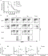
Figure 5.. Both TN and TCM expand in syngeneic and allogeneic recipients and produce IFN-γ.
BALB/c (CD45.2) and B6 (CD45.2) mice were irradiated and reconstituted with TCD B6 CD45.2 BM and 106 CD8+CD45.1+ TN or TCM. Seven days post BMT, spleen and LN cells were stimulated with PMA and ionomycin and then stained for CD45.1, CD8, IFN-γ and TNF-α. Positive gates for IFN-γ were based on isotype staining (not shown). A and B, representative flow cytometry of CD8+CD45.1+ splenocytes and LN cells, respectively. The percentage of IFN-γ+ and IFN-γ very bright cells are shown in the upper left and upper right corners, respectively. Total numbers of CD45.1+CD8+, CD45.1+CD8+IFN-γ+ or IFN-γ-bright and the percent of CD8+CD45.1+ cells that are IFN-γ+ is shown for spleen (C) and LN (D). Each circle is the value from an individual mouse; horizontal lines are mean values. E, IFN-γ expression after PMA/ionomycin treatment of freshly isolated TCM (right panel) and TN (left panel).
Figure 6.
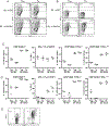
Figure 6.. Allogeneic TCM and TN induce serum IFN-γ.
Serum was harvested from B6→B6 and B6→BALB/c recipients of B6 TN or TCM on day +7. Cytokine concentrations were determined by Bio Plex beads. IFN-γ but not IL-2 or TNF-α were elevated in allogeneic TCM or TN recipients. Each symbol is a measurement from an individual mouse; horizontal lines are means.
Figure 7.
Figure 7.. Allogeneic BALB/c B cells are preferentially killed in vivo in recipients of B6 TCM or TN 7 days post transplant.
Representative flow cytometry of splenocytes (SPL) and lymph node cells (LN). Plots are gated on B220+ cells. Note that greater than 94% of CFSE+B220+ cells are H-2Kb+ and therefore B6 in recipients of TN or TCM.

References

    1. Mackall CL, and Gress RE 1997. Pathways of T-cell regeneration in mice and humans: implications for bone marrow transplantation and immunotherapy. Immunol Rev 157:61–72. - PubMed
    1. Collins RH Jr., Shpilberg O, Drobyski WR, Porter DL, Giralt S, Champlin R, Goodman SA, Wolff SN, Hu W, Verfaillie C, List A, Dalton W, Ognoskie N, Chetrit A, Antin JH, and Nemunaitis J 1997. Donor leukocyte infusions in 140 patients with relapsed malignancy after allogeneic bone marrow transplantation. J Clin Oncol 15:433–444. - PubMed (V体育2025版)
    1. Kolb HJ, Schattenberg A, Goldman JM, Hertenstein B, Jacobsen N, Arcese W, Ljungman P, Ferrant A, Verdonck L, Niederwieser D, and et al. 1995. Graft-versus-leukemia effect of donor lymphocyte transfusions in marrow grafted patients. European Group for Blood and Marrow Transplantation Working Party Chronic Leukemia. Blood 86:2041–2050. - "V体育官网入口" PubMed
    1. Porter DL, Collins RH Jr., Shpilberg O, Drobyski WR, Connors JM, Sproles A, and Antin JH 1999. Long-term follow-up of patients who achieved complete remission after donor leukocyte infusions. Biol Blood Marrow Transplant 5:253–261. - V体育安卓版 - PubMed
    1. Horowitz MM, Gale RP, Sondel PM, Goldman JM, Kersey J, Kolb HJ, Rimm AA, Ringden O, Rozman C, Speck B, and et al. 1990. Graft-versus-leukemia reactions after bone marrow transplantation. Blood 75:555–562. - "V体育安卓版" PubMed

V体育2025版 - Publication types

MeSH terms