Skip to main page content
U.S. flag

An official website of the United States government

Dot gov

The . gov means it’s official. Federal government websites often end in . gov or . mil VSports app下载. Before sharing sensitive information, make sure you’re on a federal government site. .

Https

The site is secure. The https:// ensures that you are connecting to the official website and that any information you provide is encrypted and transmitted securely. V体育官网.

Comparative Study
. 2009 Jul;33(7):1220-30.
doi: 10.1111/j.1530-0277.2009.00946.x. Epub 2009 Apr 9.

Nitric oxide-mediated intestinal injury is required for alcohol-induced gut leakiness and liver damage

Affiliations
Comparative Study

Nitric oxide-mediated intestinal injury is required for alcohol-induced gut leakiness and liver damage (VSports最新版本)

Yueming Tang (VSports在线直播) et al. Alcohol Clin Exp Res. 2009 Jul.

Abstract

Background: Alcoholic liver disease (ALD) requires endotoxemia and is commonly associated with intestinal barrier leakiness. Using monolayers of intestinal epithelial cells as an in vitro barrier model, we showed that ethanol-induced intestinal barrier disruption is mediated by inducible nitric oxide synthase (iNOS) upregulation, nitric oxide (NO) overproduction, and oxidation/nitration of cytoskeletal proteins. We hypothesized that iNOS inhibitors [NG-nitro-l-arginine methyl ester (l-NAME), l-N(6)-(1-iminoethyl)-lysine (l-NIL)] in vivo will inhibit the above cascade and liver injury in an animal model of alcoholic steatohepatitis (ASH). VSports手机版.

Methods: Male Sprague-Dawley rats were gavaged daily with alcohol (6 g/kg/d) or dextrose for 10 weeks +/- l-NAME, l-NIL, or vehicle. Systemic and intestinal NO levels were measured by nitrites and nitrates in urine and tissue samples, oxidative damage to the intestinal mucosa by protein carbonyl and nitrotyrosine, intestinal permeability by urinary sugar tests, and liver injury by histological inflammation scores, liver fat, and myeloperoxidase activity. V体育安卓版.

Results: Alcohol caused tissue oxidation, gut leakiness, endotoxemia, and ASH. l-NIL and l-NAME, but not the d-enantiomers, attenuated all steps in the alcohol-induced cascade including NO overproduction, oxidative tissue damage, gut leakiness, endotoxemia, hepatic inflammation, and liver injury. V体育ios版.

Conclusions: The mechanism we reported for alcohol-induced intestinal barrier disruption in vitro - NO overproduction, oxidative tissue damage, leaky gut, endotoxemia, and liver injury - appears to be relevant in vivo in an animal model of alcohol-induced liver injury. That iNOS inhibitors attenuated all steps of this cascade suggests that prevention of this cascade in alcoholics will protect the liver against the injurious effects of chronic alcohol and that iNOS may be a useful target for prevention of ALD. VSports最新版本.


Figures
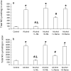
Fig. 1
Fig. 1
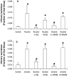
iNOS inhibitors prevented EtOH-induced NO overproduction in urine and colon mucosa. Total NO (NO2 + NO3) in urine and colonic mucosa from dextrose fed (control) and alcohol fed (10 weeks) rats was assayed after sacrifice (see Methods). Data are shown as mean for total NO (umol/mg) ± SE for n=6 for each group. The difference between groups was analyzed using ANOVA, *: p<0.05 compared to dextrose fed rats (controls), #: p<0.05 compared to alcohol fed rats, &: p<0.05 compared to alcohol + L-NAME group.
Fig. 2
Fig. 2
iNOS inhibitors prevented EtOH-induced increases in nitration (protein-nitrotyrosination) in intestinal tissues. Levels of nitrotyrosine epitopes, a marker of tissue nitration, were determined by slot blotting and quantitative densitometry in tissue (mucosal) samples obtained after sacrifice from duodenum (a), jejunum (b), ileum (c), and colon (d). Data are shown as the mean ± SE for each group (n=6/group). The difference between groups was analyzed using ANOVA, *: p<0.05 compared to dextrose-fed rats (controls),, #: p<0.05 compared to alcohol fed rats (10 weeks). A representative slot blot image for colonic mucosa is shown in Fig. 2d.
Fig. 3
Fig. 3
iNOS inhibitors prevented EtOH-induced increases in oxidation (protein-carbonyls) in intestinal tissues. Levels of the carbonyl epitope, an oxidative stress marker, were determined by slot blots and quantitative densitometry in tissue (mucosal) samples obtained after sacrifice from duodenum (a), jejunum (b), ileum (c), and colon (d). Data are shown as the mean ± SE for each group (n=6). The difference between groups was analyzed using ANOVA, *: p<0.05 compared to dextrose-fed rats (controls), #: p<0.05 compared to alcohol-fed rats (10 weeks). A representative slot blot image for colonic mucosa is shown in Fig. 3d.
Fig. 4
Fig. 4
iNOS inhibitors prevented EtOH-induced increases in intestinal permeability. Intestinal permeability was assayed by measuring urinary lactulose and sucralose after an oral dose of these sugars. Urinary lactulose (a marker of small intestine permeability and urinary sucralose (a marker of whole gut permeability) increased in alcohol fed rats. iNOS inhibitors (L-NIL or L-NAME) significantly prevented EtOH-induced hyperpermeability. Data are means of the fraction of the oral dose recovered ± S.E. for N = 6 rats for each group. The difference between groups was analyzed by ANOVA, *: p<0.05 compared to dextrose-fed rats (controls), #: p<0.05 compared to alcohol fed rats (10 weeks).
Fig. 5
Fig. 5
iNOS inhibitors prevented EtOH-induced increases in serum endotoxin. Serum endotoxin was determined for control and alcohol-fed rats at 10 weeks after alcohol feeding as described in Methods. Serum endotoxin levels were significantly higher in alcohol-fed rats. iNOS inhibitors (L-NIL or L-NAME) prevented EtOH-induced increases in endotoxin levels. Data are expressed as mean endotoxin Units (EU) per ml serum ± S.E. for N=6 rats for each group. The difference between groups was analyzed by ANOVA, *: p<0.05 compared to the control group, #: p<0.05 compared to alcohol-fed rats (10 weeks), &: p<0.05 compared to alcohol + L-NAME group.
Fig. 6
Fig. 6
iNOS inhibitors prevented EtOH-induced steatohepatitis. EtOH-induced inflammatory reactions were determined by measuring hepatic myeloperoxidase (MPO) activity (a) and by calculating histological inflammatory scores (b). Total histological score represents a combined inflammatory score (i.e. hepatitis) and score for the severity of fat accumulation in the liver (i.e. steatosis) and thus it represents the severity of steatohepatitis (c). EtOH-induced fatty liver was determined by measuring hepatic fat content (d). Feeding of alcohol to rats for 10 weeks significantly increased MPO activity, inflammation score, histology score, and fat content in liver tissue. iNOS inhibitors (L-NIL or L-NAME) significantly decreased all indices of alcoholic steatohepatitis. Data are expressed as mean ± S.E. for N=6 rats for each group. The difference between groups was analyzed using ANOVA, *: p<0.05 compared to dextrose-fed rats (controls), #: p<0.05 compared to alcohol-fed rats (10 weeks).
Fig. 7
Fig. 7
iNOS inhibitors prevented EtOH-induced increases in alanine aminotransferase (ALT). Chronic EtOH consumption by rats significantly increased serum ALT levels, an index of liver cell injury. iNOS inhibitors significantly inhibited EtOH-induced increases in ALT: back to 91+15 U/dl by L-NIL and 96+16 U/dl by L-NAME. Data are expressed as mean ± S.E. for N=6 rats for each group. Differences between groups were analyzed using ANOVA, *: p<0.05 compared to dextrose-fed rats (controls),, #: p<0.05 compared to alcohol-fed rats (10 weeks).
Fig. 8
Fig. 8
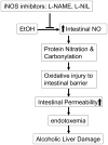
Our current model for the mechanism by which iNOS inhibitors reduce EtOH-induced steatohepatitis. They do so by preventing oxidative stress-induced increases in intestinal hyperpermeability and the endotoxemia that would otherwise result.

References

    1. Alican I, Kubes P. A critical role for nitric oxide in intestinal barrier function and dysfunction. Am J Physiol. 1996;270(2 Pt 1):G225–37. - PubMed
    1. Aram G, Potter JJ, Liu X, Torbenson MS, Mezey E. Lack of inducible nitric oxide synthase leads to increased hepatic apoptosis and decreased fibrosis in mice after chronic carbon tetrachloride administration. Hepatology. 2008;47(6):2051–8. - "V体育官网" PubMed
    1. Banan A, Choudhary S, Zhang Y, Fields JZ, Keshavarzian A. Ethanol-induced barrier dysfunction and its prevention by growth factors in human intestinal monolayers: evidence for oxidative and cytoskeletal mechanisms. J Pharmacol Exp Ther. 1999;291(3):1075–85. - PubMed
    1. Banan A, Fields JZ, Decker H, Zhang Y, Keshavarzian A. Nitric oxide and its metabolites mediate ethanol-induced microtubule disruption and intestinal barrier dysfunction. J Pharmacol Exp Ther. 2000;294(3):997–1008. - PubMed
    1. Banan A, Fields JZ, Zhang Y, Keshavarzian A. iNOS upregulation mediates oxidant-induced disruption of F-actin and barrier of intestinal monolayers. Am J Physiol Gastrointest Liver Physiol. 2001;280(6):G1234–46. - "VSports注册入口" PubMed

MeSH terms