Skip to main page content (VSports app下载)
U.S. flag

An official website of the United States government

Dot gov

The . gov means it’s official. Federal government websites often end in . gov or . mil. Before sharing sensitive information, make sure you’re on a federal government site VSports app下载. .

Https

The site is secure. The https:// ensures that you are connecting to the official website and that any information you provide is encrypted and transmitted securely V体育官网. .

. 2009 Jun;29(12):3379-89.
doi: 10.1128/MCB.01758-08. Epub 2009 Apr 13.

Extracellular signal-regulated kinase 2-dependent phosphorylation induces cytoplasmic localization and degradation of p21Cip1

Affiliations

V体育官网入口 - Extracellular signal-regulated kinase 2-dependent phosphorylation induces cytoplasmic localization and degradation of p21Cip1

Chae Young Hwang et al. Mol Cell Biol. 2009 Jun.

Abstract (V体育官网)

p21(Cip1) is an inhibitor of cell cycle progression that promotes G(1)-phase arrest by direct binding to cyclin-dependent kinase and proliferating cell nuclear antigen. Here we demonstrate that mitogenic stimuli, such as epidermal growth factor treatment and oncogenic Ras transformation, induce p21(Cip1) downregulation at the posttranslational level. This downregulation requires the sustained activation of extracellular signal-regulated kinase 2 (ERK2), which directly interacts with and phosphorylates p21(Cip1), promoting p21(Cip1) nucleocytoplasmic translocation and ubiquitin-dependent degradation, thereby resulting in cell cycle progression. ERK1 is not likely involved in this process. Phosphopeptide analysis of in vitro ERK2-phosphorylated p21(Cip1) revealed two phosphorylation sites, Thr57 and Ser130 VSports手机版. Double mutation of these sites abolished ERK2-mediated p21(Cip1) translocation and degradation, thereby impairing ERK2-dependent cell cycle progression at the G(1)/S transition. These results indicate that ERK2 activation transduces mitogenic signals, at least in part, by downregulating the cell cycle inhibitory protein p21(Cip1). .


Figures

FIG. 1.
FIG. 1.
Downregulation of p21Cip1 by ERK activation. (A) p21Cip1 levels are decreased in Ras transgenic mice. Hepatic tissue from normal and H-RasV12-transgenic mice was lysed and immunoblotted with anti-p21Cip1 and anti-phospho-ERK antibodies and normalized with anti-β-actin antibody. (B) Ras overexpression reduces p21Cip1 level. H-Ras-transformed and mock-transformed NIH 3T3 cells were lysed and immunoblotted with anti-p21Cip1, anti-phospho-ERK, and anti-H-Ras antibodies and normalized with anti-β-actin antibody. (C) ERK inhibition blocks EGF stimulation-induced p21Cip1 downregulation. HeLa cells were pretreated with U0126 (20 μM, 1 h) or LY294002 (5 μM, 1 h) and then incubated with or without EGF. Cell lysates were prepared 40 min after stimulation, analyzed by immunoblotting with anti-p21Cip1 and anti-phospho-ERK antibodies, and normalized with anti-β-actin antibody. (D and E) ERK inhibition prolongs p21Cip1 half-life. HeLa cells were pretreated with U0126 (10 μM) or dimethyl sulfoxide (vehicle) for 1 h and were incubated with cycloheximide (CHX) (10 μg/ml) in the absence or presence of EGF (50 ng/ml). At the indicated times, cell lysates were prepared, analyzed by immunoblotting with anti-p21Cip1 antibody, and normalized with anti-β-actin antibody. A representative Western blot (D) was quantified by densitometry (E). All signals are normalized to the signal at time zero (100%).
FIG. 2.
FIG. 2.
ERK2 induces proteasome-dependent p21Cip1 degradation. (A and B) ERK2 expression decreases the p21Cip1 level, an effect that is blocked by the proteasome inhibitor, MG-132. HeLa cells were transfected with a plasmid encoding FLAG-ERK2 (A) or FLAG-ERK1 (B). After 36 h, cells were incubated with or without MG132 for 4 h prior to preparing cell extracts. Extracts were analyzed by immunoblotting (I/B) with anti-p21Cip1, anti-phospho-ERK1/2 (pERK1/2), anti-FLAG (α-FLAG), anti-ERK1/2 (α-ERK1/2), and anti-β-actin antibodies. (C) DN MEK increases p21Cip1 levels. HEK293T cells were cotransfected with HA-tagged p21Cip1-encoding and increasing amounts of DN-MEK-encoding plasmids. After 40 h, cells were harvested and lysed. Extracts were analyzed by immunoblotting with anti-HA (α-HA), anti-phospho-ERK1/2, anti-phospho-MEK1/2 (pMEK), anti-MEK1/2, and anti-β-actin antibodies. (D) shRNA-mediated ERK2 knockdown increases p21Cip1 levels. HeLa cells were transfected with an ERK2 shRNA (shERK2) expression plasmid or control vector. After 48 h, cells were harvested and lysed. Extracts were analyzed by immunoblotting with anti-p21Cip1, anti-ERK2, and anti-β-actin antibodies. (E) ERK2 expression induces p21Cip1 ubiquitination. HEK293 cells were cotransfected with HA-tagged p21Cip1- and His6-tagged ubiquitin-encoding plasmids together with a FLAG-ERK2 expression plasmid or control vector. After 36 h, cells were incubated with MG132 for 4 h prior to preparation of cell extracts. His6-ubiquitin-conjugated HA-p21Cip1 proteins were purified by nickel affinity chromatography and analyzed by immunoblotting with an anti-HA antibody. Total cell extracts were analyzed by immunoblotting with anti-phospho-ERK1/2, anti-HA, anti-FLAG, anti-ERK1/2, and antiubiquitin antibodies.
FIG. 3.
FIG. 3.
p21Cip1 interacts with ERK2. (A) HEK293T cells were cotransfected with HA-p21Cip1 and FLAG-ERK2 or control vector. After 36 h, cells were lysed and ERK2 was immunoprecipitated (I/P) with anti-FLAG (α-FLAG) antibody. p21Cip1 and ERK2 were detected by immunoblotting with anti-HA (α-HA) and anti-FLAG antibodies, respectively. (B) HeLa cells were pretreated with MG132 alone and along with U0126 (10 μM) for 2 h and were treated with EGF (50 ng/ml) or left untreated for 30 min. Cells were lysed, and p21Cip1 was immunoprecipitated (I/P) with an anti-p21Cip1 (α-p21Cip1) antibody. ERK1/2, polyubiquitinated p21Cip1, and native p21Cip1 were detected by immunoblotting with anti-ERK1/2, antiubiquitin (α-ubiquitin), and anti-p21Cip1 antibodies, respectively. Total cell extracts were analyzed by immunoblotting with anti-phospho-ERK1/2, anti-ERK1/2, and anti-p21Cip1 antibodies. (C) Aligned amino acid sequences of the ERK-docking FXF motifs of human p21Cip1 and other known ERK substrates: human mitogen-activated protein kinase phosphatase 1 (hMKP1), Elk-1, phosphodiesterase 4A (PDE4A), serum response factor accessory protein 1 (hSAP-1), and mouse kinase suppressor of Ras 1 (mKsr1). Conserved residues are highlighted. Numbers on the right indicate the positions of the final residues shown in each case. (D) Aligned amino acid sequences of the ERK-docking KIM motifs of human p21Cip1 and other known ERK substrates: hMKP2, Elk-1, PDE4A, striatally enriched tyrosine phosphatase (STEP), and p90 ribosomal S6 kinase 1 (RSK1). (E) HEK293T cells were cotransfected with either HA-tagged wild-type (WT) p21Cip1 (1 μg) or docking mutant (DM) p21Cip1 (0.4 μg) and FLAG-ERK2 or control vector. Different amounts of p21Cip1 constructs were used to adjust the levels of protein expression equally. After 36 h, cells were incubated with MG132 for 4 h prior to preparing cell extracts. p21Cip1 was immunoprecipitated with an anti-HA antibody. p21Cip1 and ERK2 were detected by immunoblotting with anti-HA and anti-FLAG antibodies, respectively.
FIG. 4.
FIG. 4.
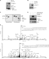
ERK2 phosphorylates Thr57 and Ser130 residues in p21Cip1 in vitro. (A) HEK293T cells transfected with either HA-p21Cip1 or HA-ERK2 were lysed and separately immunoprecipitated with anti-HA antibody. p21Cip1 and ERK2 immunoprecipitates were incubated in kinase reaction buffer containing [γ-32P]ATP. Phosphorylated proteins were resolved by SDS-PAGE and analyzed by autoradiography. The lower two panels show immunoblots (I/B) for the p21Cip1 and ERK2 proteins used in the kinase reactions, detected with an anti-HA (α-HA) antibody. (B) Recombinant ERK2 and p21Cip1 proteins were purified from E. coli and incubated in kinase reaction buffer containing [γ-32P]ATP. Phosphorylated proteins were resolved by SDS-PAGE and analyzed by autoradiography. The lower panel shows an immunoblot with anti-p21Cip1 antibody as a loading control. (C) The samples in panel B resolved by SDS-PAGE were transferred onto a polyvinylidene difluoride membrane. Radioactive protein was cut from the membrane, acid hydrolyzed, mixed with cold phosphoserine, phosphothreonine, and phosphotyrosine standards, and separated on cellulose-thin-layer chromatography plates (Merck) in two dimensions using an HTLE 7000 (CBS Scientific) apparatus. The positions of standard phosphoamino acids were determined by ninhydrin staining (left panel), and 32P-amino acids were located by autoradiography (right panel). (D) Identification of the ERK-mediated phosphorylation sites in p21Cip1. Recombinant p21Cip1 protein was phosphorylated in vitro with ERK2. After the sample was resolved on SDS-PAGE, the p21Cip1 protein band was excised and subjected to proteolysis with trypsin, and the resulting peptides were analyzed by LC-MS/MS. Two different phosphopeptides were detected. The presence of y- and b-type fragment ions in MS/MS spectra enabled identification of the tryptic peptides, SGEQAEGpSPGGPGDSQGR and ERWNFDFVTEpTPLEGDFAWER; phosphorylated Thr57 and Ser130 are indicated. (E) HEK293T cells transfected with either HA-tagged wild-type or T57A S130A p21Cip1 were lysed and immunoprecipitated with anti-HA antibody. p21Cip1 immunoprecipitates were incubated with recombinant active ERK2 (rec-ERK2) in kinase reaction buffer containing [γ-32P]ATP. Phosphorylated proteins were resolved by SDS-PAGE and analyzed by autoradiography. The lower two panels show immunoblots for the p21Cip1 and ERK2 proteins used in the kinase reactions, detected with anti-HA and anti-ERK2 antibodies, respectively.
FIG. 5.
FIG. 5.
ERK2 alters the cellular localization of p21Cip1. (A and B) HeLa cells were cotransfected with HA-tagged wild-type p21Cip1 (WT) or T57A S130A mutant p21Cip1 together with FLAG-ERK2 or the control vector. At 42 h after transfection, cells were treated with MG132 for 2 h prior to fixation. The cellular localization of p21Cip1 was detected using an anti-HA antibody. After washing extensively in PBS, samples were further incubated with an anti-FLAG antibody to detect ERK2. Nuclei were visualized by DAPI staining. Cells were observed by fluorescence microscopy. A representative microscopic field in the lower middle panel in A shows that nuclear and cytoplasmic localization of p21Cip1 is indicated by two cells (arrows) coexpressing FLAG-ERK2 (red signals in ERK2 staining; right panel) and HA-p21Cip1 and that nuclear localization of p21Cip1 is indicated by two cells (starred) expressing HA-p21Cip1 only (no signal in ERK2 staining; right panel). (C) HeLa cells were transfected with HA-tagged wild-type p21Cip1. At 42 h after transfection, cells were treated with MG132 and U0126 (10 μM) or DMSO (vehicle) for 2 h prior to treatment with EGF (50 ng/ml) for 30 min. After fixation, the cellular localization of p21Cip1 was detected using an anti-HA antibody. Nuclei were visualized by DAPI staining. Cells were observed by fluorescence microscopy. (D) Cells in A and B were fractionated, and cytoplasmic (C) and nuclear (N) proteins were resolved by SDS-PAGE and analyzed by immunoblotting with anti-HA (α-HA), anti-FLAG (α-FLAG), antitubulin, and anti-lamin A/C antibodies. Lamin A/C and tubulin are nuclear and cytoplasmic markers, respectively. (E) HeLa cells were treated with MG132 and U0126 (10 μM) or with DMSO (vehicle) for 2 h prior to treatment with EGF (50 ng/ml) for 30 min. Cytoplasmic (C) and nuclear (N) proteins were resolved by SDS-PAGE and analyzed by immunoblotting with anti-p21Cip1, anti-pERK1/2, anti-ERK1/2, antitubulin, and anti-PARP-1 antibodies. PARP-1 and tubulin are nuclear and cytoplasmic markers, respectively.
FIG. 6.
FIG. 6.
ERK2 phosphorylation of p21Cip1 at Thr57 and Ser130 induces p21Cip1 ubiquitination and subsequent degradation. (A) HEK293 cells were cotransfected with HA-tagged wild-type (WT) p21Cip1, T57A S130A mutant p21Cip1, or docking mutant (DM) p21Cip1 together with His6-ubiquitin and FLAG-ERK2 or a control vector. After 36 h, cells were incubated with MG132 for 4 h prior to preparation of cell extracts. His6-ubiquitin conjugates were purified by nickel affinity chromatography and analyzed by immunoblotting (I/B) with an anti-HA (α-HA) antibody. Total cell extracts were analyzed by immunoblotting with anti-HA, anti-FLAG (α-FLAG), and antiubiquitin antibodies. (B) HEK293 cells were cotransfected with either HA-tagged wild-type (WT) p21Cip1 or T57A S130A mutant p21Cip1 and FLAG-ERK2 or control vector. Cells were cotransfected with an enhanced-GFP vector for normalization. After 40 h, cells were harvested and lysed, and extracts were analyzed by immunoblotting with anti-HA, anti-phospho-ERK1/2, anti-FLAG, and anti-GFP antibodies. (C) HeLa cells were transfected with HA-tagged docking mutant p21Cip1 together with FLAG-ERK2 or a control vector. After 48 h, cells were harvested, and cell extracts were analyzed by immunoblotting with anti-p21Cip1 (α-p21Cip1), anti-FLAG, and anti-β-actin antibodies.
FIG. 7.
FIG. 7.
ERK2 promotes cell cycle progression via degradation of p21Cip1. (A) HEK293 cells were transfected with HA-tagged wild-type (WT) p21Cip1 or T57A S130A mutant p21Cip1 together with FLAG-ERK2 or a control vector. A vector encoding membrane-bound GFP was cotransfected, and cells were incubated for 32 h. During the last 16 h, cells were starved in serum-free medium and then returned to normal medium for an additional 8 h before harvesting. The cell cycle profile of GFP-positive cells was determined by fluorescence-activated cell sorting analysis. (B) HCT116 p21−/− cells were transfected with HA-tagged wild-type (WT) p21Cip1 or T57A S130A mutant p21Cip1 together with FLAG-ERK2 or a control vector and a membrane-bound GFP vector. Cells were treated and analyzed as for panel A. The levels of HA-p21Cip1 and FLAG-ERK2 were analyzed by immunoblotting (see Fig. S2 in the supplemental material). (C) Schematic representation of the proposed pathway, showing that ERK2 interacts with and phosphorylates p21Cip1 at Thr57 and Ser130; phosphorylation of these residues enhances p21Cip1 ubiquitination and subsequent degradation through the proteasome pathway, leading to cell proliferation.

References

    1. Albanese, C., J. Johnson, G. Watanabe, N. Eklund, D. Vu, A. Arnold, and R. G. Pestell. 1995. Transforming p21ras mutants and c-Ets-2 activate the cyclin D1 promoter through distinguishable regions. J. Biol. Chem. 27023589-23597. - PubMed
    1. Bendjennat, M., J. Boulaire, T. Jascur, H. Brickner, V. Barbier, A. Sarasin, A. Fotedar, and R. Fotedar. 2003. UV irradiation triggers ubiquitin-dependent degradation of p21(WAF1) to promote DNA repair. Cell 114599-610. - PubMed
    1. Blagosklonny, M. V., G. S. Wu, S. Omura, and W. S. el-Deiry. 1996. Proteasome-dependent regulation of p21WAF1/CIP1 expression. Biochem. Biophys. Res. Commun. 227564-569. - V体育2025版 - PubMed
    1. Bloom, J., V. Amador, F. Bartolini, G. DeMartino, and M. Pagano. 2003. Proteasome-mediated degradation of p21 via N-terminal ubiquitinylation. Cell 11571-82. - PubMed
    1. Bornstein, G., J. Bloom, D. Sitry-Shevah, K. Nakayama, M. Pagano, and A. Hershko. 2003. Role of the SCFSkp2 ubiquitin ligase in the degradation of p21Cip1 in S phase. J. Biol. Chem. 27825752-25757. - PubMed

Publication types

VSports app下载 - MeSH terms