Skip to main page content
U.S. flag

An official website of the United States government

Dot gov

The . gov means it’s official VSports app下载. Federal government websites often end in . gov or . mil. Before sharing sensitive information, make sure you’re on a federal government site. .

Https

The site is secure V体育官网. The https:// ensures that you are connecting to the official website and that any information you provide is encrypted and transmitted securely. .

. 2009 Apr 13;206(4):793-805.
doi: 10.1084/jem.20081648. Epub 2009 Mar 16.

Normally occurring NKG2D+CD4+ T cells are immunosuppressive and inversely correlated with disease activity in juvenile-onset lupus

Affiliations

Normally occurring NKG2D+CD4+ T cells are immunosuppressive and inversely correlated with disease activity in juvenile-onset lupus (V体育官网)

Zhenpeng Dai (VSports app下载) et al. J Exp Med. .

"VSports最新版本" Abstract

The NKG2D receptor stimulates natural killer cell and T cell responses upon engagement of ligands associated with malignancies and certain autoimmune diseases. However, conditions of persistent NKG2D ligand expression can lead to immunosuppression. In cancer patients, tumor expression and shedding of the MHC class I-related chain A (MICA) ligand of NKG2D drives proliferative expansions of NKG2D(+)CD4(+) T cells that produce interleukin-10 (IL-10) and transforming growth factor-beta, as well as Fas ligand, which inhibits bystander T cell proliferation in vitro. Here, we show that increased frequencies of functionally equivalent NKG2D(+)CD4(+) T cells are inversely correlated with disease activity in juvenile-onset systemic lupus erythematosus (SLE), suggesting that these T cells may have regulatory effects VSports手机版. The NKG2D(+)CD4(+) T cells correspond to a normally occurring small CD4 T cell subset that is autoreactive, primed to produce IL-10, and clearly distinct from proinflammatory and cytolytic CD4 T cells with cytokine-induced NKG2D expression that occur in rheumatoid arthritis and Crohn's disease. As classical regulatory T cell functions are typically impaired in SLE, it may be clinically significant that the immunosuppressive NKG2D(+)CD4(+) T cells appear functionally uncompromised in this disease. .


Figures
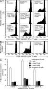
Figure 1.
Figure 1.
Autoreactivity of normal peripheral blood NKG2D+CD4+ T cells. (A) NKG2D+CD4+ T cells proliferate in the presence of CD25+ cell–depleted autologous PBMCs. CD3+CD4+CD8NKG2D+ T cells FACSAria-sorted from magnetic bead–enriched CD4+ T cells to ≥99% purity were labeled with CFSE and cultured with irradiated autologous total or CD25 stimulator PBMCs, or subpopulations thereof, as indicated. CD25+ lymphocytes were depleted to enhance T cell proliferation (23). Autoreactive NKG2D+CD4+ T cell stimulation is solely dependent on B cells as shown separately by B cell depletion and reconstitution. T cell and monocyte depletions have no effect. Blocking antibodies are specific for MHC class II (mAb TU39), and ULBP2 or ULBP3 (mAbs 6F6 and 4F9). The CFSE profiles shown represent gated NKG2D+CD4+ T cells. (B) Purified NKG2DCD4+ T cells fail to proliferate in the presence of CD25+ cell–depleted autologous PBMCs. Treatment of NKG2DCD4+ T cells with PHA is shown as positive control. CFSE profiles represent gated NKG2DCD4+ T cells. Data shown in A and B are representative of results obtained in duplicate experiments with PBMCs from five independent donors. (C) Thymidine incorporation by NKG2D+CD4+ T cell clones in the presence of autologous, but not allogeneic, B cells is inhibited by anti-MHC class II (mAb TU39) and -HLA-DR (mAb L243). Clones 1–3 were derived from 3 donors and are representative of a total of 32 clones. Error bars indicate deviations among triplicate wells. Data shown are representative of three experiments per clone.
Figure 2.
Figure 2.
Autoreactivity of phenotypically naive NKG2D+CD4+ T cells. (A) CD3+CD4+CD8CD45RA+CD45ROCD62L+CCR7+CD28+CD27+NKG2D+ T cells FACSAria sorted from magnetic bead–enriched CD4+ T cells to >99% purity dilute CFSE in the presence of autologous, but not allogeneic, B cells. Proliferation is inhibited by anti-MHC class II mAb TU39. CFSE profiles shown represent gated NKG2D+CD4+ T cells and are representative of data obtained in duplicate experiments with PBMCs from five independent donors. (B) NKG2D+CD4+ T cell clones derived from single-cell sorted naive NKG2D+CD4+ T cells incorporate [3H]thymidine in the presence of autologous, but not allogeneic B cells. Proliferative responses are inhibited by mAb TU39. Clones 1–3 are representative of a total of 60 clones established from 3 donors. Error bars indicate deviations among triplicate wells. Data shown are representative of three experiments per clone.
Figure 3.
Figure 3.
Absence of recall responses by NKG2D+CD4+ T cells. (A) By CFSE dilution, purified memory NKG2D+CD8CD4+ T cells are unresponsive to stimulation by autologous monocytes pulsed with CMV pp65, tetanus toxoid (TT), M. tuberculosis–purified protein derivative (Mt PPD), and C. albicans (Ca) antigen cocktail. (B) Proliferation of NKG2D+CD4+ T cells nondepleted for CD8 coexpressing T cells. (C) CFSE diluting T cells seen in B (profile left of vertical line) are double-positive CD8αβ+CD4+ NKG2D+ T cells. (D−G) Proliferation of donor-matched purified memory NKG2DCD4+ T cells upon stimulation by monocytes pulsed with the individual recall antigens. Data shown are representative of results obtained in duplicate experiments with PBMC from five donors.
Figure 4.
Figure 4.
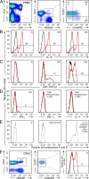
IL-10 and TGF-β cytokine signature of peripheral blood and cord blood NKG2D+CD4+ T cells after short-term stimulation with PMA and ionomycin. (A) Polychromatic flow cytometry gating tree for naive (N; CD45ROCCR7+), central memory (CM; CD45RO+CCR7+), and effector memory (EM; CD45ROCCR7) NKG2D+CD8CD4+CD3+ T cells. Numbers in quadrants and square gate specify T cell frequencies (in percent). All histograms shown in B–D were generated based on this gating tree. (B) Staining for intracellular IL-10 of naive (N) and central (CM) and effector (EM) memory NKG2D+CD4+ T cells. (C) Staining of surface LAP-TGF-β1 on memory (CM and EM), but not naive (N), NKG2D+CD4+ T cells. (D) Minimal detection of intracellular IFN-γ and absence of TNF-α and IL-17 in effector memory (EM) and total NKG2D+CD4+ T cell populations, respectively. (E) Lack of intracellular staining of naive and memory (CM and EM) NKG2D+CD4+ T cells for perforin and granzymes A and B using an antibody cocktail. Gating was as shown in A. Histogram at right shows positive control staining of CD4 T cells with IL-15–induced NKG2D expression (11). (F) Gating tree and intracellular IL-10 staining of naive (CD45ROCCR7+) NKG2D+CD4+ T cells among CBMCs. Numbers specify T cell frequencies (in percent). Numbers above horizontal bars in histograms B−D and F indicate the percentage of positive cells. Histograms shown in red and black are from stimulated and unstimulated T cells, respectively. Data shown are representative of results obtained in duplicate experiments with samples from 10 donors.
Figure 5.
Figure 5.
IL-10, TGF-β, IFN-γ, and FasL production by normal peripheral blood-derived NKG2D+CD4+ T cell clones. (A) Detection of intracellular IL-10 and surface LAP-TGF-β1, and of IL-10 and IFN-γ among clones established under nonpolarizing conditions from sorted naive (24 T cell clones) and central and effector (CM and EM) memory (30 T cell clones) and effector (CM) memory (20 T cell clones) NKG2D+CD4+ T cells, respectively. Dots in scatter plots represent the percentage of positive cells among a given T cell clone. Clones were derived from three donors. (B) ELISA for FasL in supernatants of NKG2D+CD4+ T cell clones (identical to those used in Fig. 1) generated by autologous B cell stimulation. Conditions of experimental T cell stimulations are indicated in the bar graph. Data in A and B are representative of three separate experiments.
Figure 6.
Figure 6.
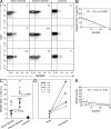
Increased proportions of NKG2D+CD4+ T cells and inverse correlation with disease activity in juvenile-onset SLE. (A) Frequencies of NKG2D+CD4+ T cells (percentage of CD8CD4+ T cells) in six disease and three age-matched control samples. (B) Overall data point distribution and statistical evaluation (R = −0.5; P = 0.02) of NKG2D+CD4+ T cell frequencies in relationship to SLEDAI scores in the 27 patient samples studied. (C) Comparison of mean frequencies (positions indicated by short horizontal bars) of NKG2D+CD4+ T cells in 13 active and 14 inactive juvenile-onset SLE and 14 age-matched control samples. Individual comparisons are bracketed, and P values are indicated. (D) NKG2D+CD4+ T cell frequencies in five patients at times of flare or remission of disease. Numbers refer to SLE subjects listed in Table I. (E) Plotting of sMICA concentrations in 72 juvenile-onset SLE plasma samples against SLEDAI scores (R = −0.27; P = 0.01). All data shown in A–E were derived from single analyses of patient and control samples.
Figure 7.
Figure 7.
Autoreactivity and cytokine signature of NKG2D+CD4+ T cells in juvenile-onset SLE. (A) Left histogram shows CFSE dilution of sorted patient-derived NKG2D+CD4+ T cells in response to stimulation by autologous (open profile), but not allogeneic B cells (shaded profile). Data shown are representative of results obtained from single analyses of two samples each from patients with active or inactive disease. Middle and right histograms show stainings for intracellular IL-10 and surface LAP-TGF-b1 of patient NKG2D+CD4+ T cells stimulated with PMA and ionomycin. Data shown are representative of results obtained with 13 active and 14 inactive disease samples analyzed once. (B) Minimal staining for intracellular IFN-γ, and absence of staining for TNF-α and IL-17 among effector memory (EM) and total patient NKG2D+CD4+ T cell populations, respectively. Data shown are representative of results obtained with 13 active and 14 inactive disease samples analyzed once. All histograms displaying intracellular cytokine stainings were generated based on the gating tree shown in Fig. 4 A. Numbers above horizontal bars in histograms indicate the percentage of positive cells. Histograms shown in black and gray are from stimulated and unstimulated T cells, respectively.
Figure 8.
Figure 8.
Functional competence of NKG2D+CD4+ T cells in juvenile-onset SLE. (A) IL-10, TGF-β, and FasL in supernatants of NKG2D+CD4+ T cells from active and inactive disease patients and age-matched controls. Sorted T cells were stimulated as indicated, and mediators were determined by ELISA. Data shown represent means of values obtained with each of five active and inactive SLE patient and control samples that were analyzed once. Error bars indicate SDs. (B and C) CFSE dilution histograms of gated NKG2DCD4+ T cells (B, normal donor T cell line; C, ex vivo–sorted patient T cells) co-cultured with purified patient or control NKG2D+CD4+ T cells activated with solid-phase anti-CD3 alone and exposed to sMICA alone or together with the antagonist anti-Fas. Data shown are representative of each of five active and inactive SLE patient and control samples analyzed once. (D) Plotting of IL-10 and FasL plasma concentrations against NKG2D+CD4+ T cell frequencies (R = 0.85, P < 0.0001; and R = 0.68, P = 0.0003) from 27 juvenile-onset SLE patient samples analyzed once.

References (V体育安卓版)

    1. Long E.O. 1999. Regulation of immune responses through inhibitory receptors.Annu. Rev. Immunol. 17:875–904 - PubMed
    1. Huard B., Karlsson L. 2000. KIR expression on self-reactive CD8+ T cells is controlled by T-cell receptor engagement.Nature. 403:325–328 - "V体育平台登录" PubMed
    1. Moser J.M., Gibbs J., Jensen P.E., Lukacher A.E. 2002. CD94-NKG2A receptors regulate antiviral CD8(+) T cell responses.Nat. Immunol. 3:189–195 - PubMed
    1. Lanier L.L. 2008. Up on the tightrope: natural killer cell activation and inhibition.Nat. Immunol. 9:495–502 - "V体育ios版" PMC - PubMed
    1. Groh V., Steinle A., Bauer S., Spies T. 1998. Recognition of stress-induced MHC molecules by intestinal epithelial gammadelta T cells.Science. 279:1737–1740 - PubMed

VSports注册入口 - Publication types

MeSH terms