Skip to main page content
U.S. flag

An official website of the United States government

Dot gov

The . gov means it’s official. Federal government websites often end in . gov or VSports app下载. mil. Before sharing sensitive information, make sure you’re on a federal government site. .

Https

The site is secure. The https:// ensures that you are connecting to the official website and that any information you provide is encrypted and transmitted securely V体育官网. .

. 2009 May;77(5):1734-45.
doi: 10.1128/IAI.00027-09. Epub 2009 Mar 9.

An Ehrlichia chaffeensis tandem repeat protein interacts with multiple host targets involved in cell signaling, transcriptional regulation, and vesicle trafficking

Affiliations

"V体育官网入口" An Ehrlichia chaffeensis tandem repeat protein interacts with multiple host targets involved in cell signaling, transcriptional regulation, and vesicle trafficking

Abdul Wakeel et al. Infect Immun. 2009 May.

Abstract

Ehrlichia chaffeensis is an obligately intracellular bacterium that exhibits tropism for mononuclear phagocytes forming cytoplasmic membrane-bound microcolonies called morulae. To survive and replicate within phagocytes, E. chaffeensis exploits the host cell by modulating a number of host cell processes, but the ehrlichial effector proteins involved are unknown. In this study, we determined that p47, a secreted, differentially expressed, tandem repeat (TR) protein, interacts with multiple host proteins associated with cell signaling, transcriptional regulation, and vesicle trafficking. Yeast two-hybrid analysis revealed that p47 interacts with polycomb group ring finger 5 (PCGF5) protein, Src protein tyrosine kinase FYN (FYN), protein tyrosine phosphatase non-receptor type 2 (PTPN2), and adenylate cyclase-associated protein 1 (CAP1). p47 interaction with these proteins was further confirmed by coimmunoprecipitation assays and colocalization in HeLa cells transfected with p47-green fluorescent fusion protein (AcGFP1-p47). Moreover, confocal microscopy demonstrated p47-expressing dense-cored (DC) ehrlichiae colocalized with PCGF5, FYN, PTPN2, and CAP1. An amino-terminally truncated form of p47 containing TRs interacted only with PCGF5 and not with FYN, PTPN2, and CAP1, indicating differences in p47 domains that are involved in these interactions VSports手机版. These results demonstrate that p47 is involved in a complex network of interactions involving numerous host cell proteins. Furthermore, this study provides a new insight into the molecular and functional distinction of DC ehrlichiae, as well as the effector proteins involved in facilitating ehrlichial survival in mononuclear phagocytes. .


Figures
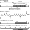
FIG. 1.
FIG. 1.
Schematic representation of E. chaffeensis p47 and regions of homology with renin receptor and DNA polymerase III subunits. (A) The amino (N)-terminal region is shaded (white bar) and precedes the seven 19-mer (ASVSEGDAVVNAVSQETPA) TRs (47% of the total protein sequence) region (gray bars). (B) The surface accessibility curve (13) predicts that each of the seven 19-mer TRs (numbered 1 to 7 over the curve) is surface exposed. (C) The C-terminal region of p47 contains six renin receptor-like motifs (14). (D) The DNA polymerase III subunit gamma and tau conserved domain (multi-domain PRK05648) was identified from NCBI Conserved Domain Database (CDD) (1). The computationally detected multi-domains are present in the same region of C-terminal region of p47 that contains the seven 19-mer TRs.
FIG. 2.
FIG. 2.
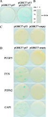
Yeast two-hybrid human bone marrow library clones that interacted with E. chaffeensis p47. (A) E. chaffeensis p47 was screened for autoactivation in S. cerevisiae strain AH109 containing pGBKT7-p47 and a positive control (AH109/Y187 diploid with pGBKT7-p53 [murine p53] and pGADT7-T [simian virus 40 large T antigen] generated by mating the S. cerevisiae strains AH109 [pGBKT7-53] and Y187 [pGADT7-T]) and selected with TDO medium. (B) Expression of p47/GAL4-BD (arrow; p47 size = 47 kDa + 21-kDa GAL4-BD tag [68 kDa total]) was determined by immunoblotting of yeast cell extracts transformed with pGBKT7-p47 (lane 1) or pGBKT7-empty (lane 2) using anti-GAL4-BD antibody. (C) To identify possible interacting clones, diploids were selected on QDO medium. A positive interaction was detected by using a diploid containing pGBKT7-p53 and pGADT7-T (plate on the left side of panel C), and the diploid containing pGBKT7-empty and pGADT7-T was used as a negative control (plate on right side of panel C). (D) S. cerevisiae strain AH109 expressing pGBKT7-p47 was mated with S. cerevisiae strain Y187 expressing PCGF5, FYN, PTPN2, and CAP1 (plates on the left side of panel D). S. cerevisiae strain AH109 expressing pGBKT7-empty was mated with S. cerevisiae strain Y187 expressing PCGF5, FYN, PTPN2, and CAP1 (plates on the right side of panel D). Diploids were selected as described for panel C.
FIG. 3.
FIG. 3.
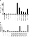
E. chaffeensis p47 interaction with multiple human proteins with distinct functional properties. AcGFP (control, without insert) and AcGFP-p47 were coexpressed with ProLabel (control, without insert) and ProLabel-PCGF5, -FYN, -PTPN2, -CAP1, -IGLL1 as fusion proteins in HeLa cells. ProLabel activity was measured at different time intervals after addition of substrate. The ProLabel tag associated with AcGFP-p47 fusion protein and enzyme acceptor combine to form an active enzyme that cleaves the chemiluminescent substrate. The resulting signal is detected with a luminometer as RLU. The ProLabel tag is the limiting constituent and is governed by its interaction with AcGFP-p47 fusion protein. (A) Relative strength and interaction of AcGFP and AcGFP-p47 with ProLabel-PCGF5, -FYN, -PTPN2, -CAP1, and -IGLL1. The results are from three independent experiments. The values are means ± standard deviations shown as RLU. An asterisk indicates that the RLU of AcGFP-p47+PCGF5, AcGFP-p47+FYN, AcGFP-p47+PTPN2, AcGFP-p47+CAP1, AcGFP-p47+IGLL1 were significantly higher than those of AcGFP+PCGF5, AcGFP+FYN, AcGFP+PTPN2, AcGFP+CAP1, and AcGFP+IGLL1 (P < 0.005). (B) Relative strength and interaction of AcGFP, AcGFP-Nterm p47, and AcGFP-Cterm p47 with PL-PCGF5, -FYN, -PTPN2, and -CAP1. The results are from three independent experiments. The values are means ± standard deviations shown as RLU. The RLU of AcGFP-Ctermp47+PCGF5 was significantly higher than those of AcGFP+PCGF5 (*, P < 0.005). AcGFP, pAcGFP1 without insert; AcGFP-p47, pAcGFP1-p47; PL-C, ProLabel without insert; PCGF5, FYN, PTPN2, CAP1, and IGLL1, PL-PCGF5, PL-FYN, PL-PTPN2, PL-CAP1, and PL-IGLL1, respectively; Nterm p47, amino-terminal p47; Cterm p47, carboxy-terminal p47.
FIG. 4.
FIG. 4.
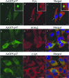
Colocalization of FYN, PTPN2, and CAP1 with AcGFP-p47 in HeLa cells. pAcGFP-p47-transfected HeLa cells (2 days posttransfection) were labeled and observed by confocal laser-scanning microscopy. The AcGFP-p47 (green) (A, D, and G) and anti-FYN (red; B), anti-PTPN2 (red; E), and anti-CAP1 (red; H) signals were merged, respectively (C, F, and I). FYN, PTPN2, and CAP1 colocalize with p47 (C, F, and I). The arrow indicates the punctulated colocalization of p47 and FYN. In the insets (A, B, and C), the arrow indicates an area highlighted to show the membrane colocalization of p47 and FYN. Small punctulated colocalization of p47 and PTPN2 is observed mostly in the cytoplasm of HeLa cells (F). Bar, 10 μm.
FIG. 5.
FIG. 5.
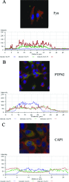
Fluorescent confocal microscopy and intensity profiles of HeLa cells expressing AcGFP-p47 (p47-green) fixed and stained with DAPI (blue) and anti-FYN, -PTPN2, or -CAP1 antibodies (red) showing colocalization of p47 with FYN (A), PTPN2 (B), and CAP1 (C). The red line in the confocal image indicates the area selected for fluorescence intensity profile analysis and is displayed in graph form (below). The x axis shows distance (μm), and the y axis shows intensity (0 to 250 arbitrary units).
FIG. 6.
FIG. 6.
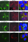
Colocalization of E. chaffeensis p47 with PCGF5, FYN, PTPN2, and CAP1 in E. chaffeensis-infected THP-1 cells. THP-1 cells were infected with E. chaffeensis and 3 days postinfection were dually labeled and examined by confocal microscopy. The panels on the right are merged images. PCGF5, FYN, PTPN2, and CAP1 colocalize with E. chaffeensis p47-labeled morulae (yellow, right panels). Differential expression of E. chaffeensis p47 by a subset of ehrlichiae compared to Dsb (constitutively expressed by E. chaffeensis) is revealed by double-immunofluorescent labeling of E. chaffeensis-infected THP-1 cells with anti-E. chaffeensis Dsb (inset in Fig. 6A, green) and anti-p47 (inset in Fig. 6B, red) sera; the merged image (inset in Fig. 6C) demonstrates the singly (green, marked with arrowhead) and dually labeled (yellow, marked with arrow) E. chaffeensis. In the inset (bottom, right panel), a normal uninfected THP-1 cell reveals that CAP1 is mainly associated with plasma membrane, while in the E. chaffeensis-infected THP-1 cell, CAP1 is distributed in cytoplasm and associated with E. chaffeensis-containing morulae. Bar, 10 μm.
FIG. 7.
FIG. 7.
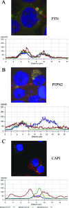
Fluorescent confocal microscopy and intensity profiles of E. chaffensis-infected THP-1 cells fixed and stained with DAPI (blue), anti-p47 (green) and anti-FYN, -PTPN2, or -CAP1 antibodies (red) showing colocalization of p47 with FYN (A), PTPN2 (B), and CAP1 (C). The red line in the confocal image indicates the area selected for fluorescence intensity profile analysis and is displayed in graph form (below). The x axis shows distance (μm), and the y axis shows the intensity (0 to 250 arbitrary units). FYN (red) and PTPN2 (red) colocalize and associate with p47 (green), as revealed in diagrams A and B, respectively. In panel C, the profile along the E. chaffeensis-infected THP-1 cells displays that CAP1 (red) surrounds the E. chaffeensis-containing morulae stained with p47 (green) and also with DAPI (blue).
FIG. 8.
FIG. 8.
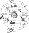
Proposed model of the role of p47 in E. chaffeensis infection and survival in monocytes based on this study and previous reports (22-24, 51, 52). Binding of E. chaffeensis to its receptor directly or indirectly activates FYN. Activated FYN tyrosine kinase phosphorylates and activates PLC-γ, which hydrolyzes membrane phospholipid PIP2 (phosphatidylinositol biphosphate), resulting in increased level of IP3 and release of Ca2+ from intracellular stores, and Ca2+ influx. FYN may regulate the function of IP3 receptor by phosphorylation and promoting release of Ca2+ from the endoplasmic reticulum. FYN also phosphorylates caveolin-1 involved in ehrlichial entry. p47-PTPN2 interaction modulates cytokine signaling events by exerting negative feedback on the Jak/Stat pathway by dephosphorylation of Jaks and Stats involved in intravacuolar maintenance and survival of E. chaffeensis. p47-PCGF5 interaction modulates gene transcription associated with cell signaling and remodeling of cytoskeleton, facilitating and supporting intracellular survival of E. chaffeensis. CAP1 promotes rapid actin dynamics in conjunction with ADF/cofilin and is required for cell morphology, migration, and endocytosis. Mitochondrial shuttling of CAP1 promotes actin- and cofilin-dependent apoptosis, which may play important role in release of DC ehrlichiae from the monocyte.

References

    1. Altschul, S. F., T. L. Madden, A. A. Schaffer, J. Zhang, Z. Zhang, W. Miller, and D. J. Lipman. 1997. Gapped BLAST and PSI-BLAST: a new generation of protein database search programs. Nucleic Acids Res. 253389-3402. - PMC (VSports app下载) - PubMed
    1. Anderson, R. G. 1998. The caveolae membrane system. Annu. Rev. Biochem. 67199-225. - PubMed
    1. Barnewall, R. E., N. Ohashi, and Y. Rikihisa. 1999. Ehrlichia chaffeensis and E. sennetsu, but not the human granulocytic ehrlichiosis agent, colocalize with transferrin receptor and up-regulate transferrin receptor mRNA by activating iron-responsive protein 1. Infect. Immun. 672258-2265. - PMC - PubMed
    1. Barnewall, R. E., Y. Rikihisa, and E. H. Lee. 1997. Ehrlichia chaffeensis inclusions are early endosomes which selectively accumulate transferrin receptor. Infect. Immun. 651455-1461. - PMC - PubMed
    1. Bertling, E., O. Quintero-Monzon, P. K. Mattila, B. L. Goode, and P. Lappalainen. 2007. Mechanism and biological role of profilin-Srv2/CAP interaction. J. Cell Sci. 1201225-1234. - PubMed

Publication types (V体育平台登录)

V体育2025版 - MeSH terms

Substances

"VSports手机版" LinkOut - more resources