Skip to main page content
U.S. flag

An official website of the United States government

Dot gov

The . gov means it’s official. Federal government websites often end in . gov or . mil. Before sharing sensitive information, make sure you’re on a federal government site VSports app下载. .

Https

The site is secure. The https:// ensures that you are connecting to the official website and that any information you provide is encrypted and transmitted securely. V体育官网.

. 2009 May;17(5):880-8.
doi: 10.1038/mt.2009.34. Epub 2009 Mar 3.

Genetic manipulation of tumor-specific cytotoxic T lymphocytes to restore responsiveness to IL-7

Affiliations

Genetic manipulation of tumor-specific cytotoxic T lymphocytes to restore responsiveness to IL-7 (V体育2025版)

Juan F Vera et al. Mol Ther. 2009 May.

Abstract

Adoptive transfer of antigen-specific cytotoxic T lymphocytes (CTLs) can induce objective clinical responses in patients with malignant diseases. The option of providing a proliferative and survival advantage to adoptively transferred CTLs remains a challenge to improve their efficacy. Host lymphodepletion and administration of recombinant interleukin-2 (IL-2) are currently used to improve CTL survival and expansion after adoptive transfer, but these approaches are frequently associated with significant side effects and may increase proliferation of T regulatory cells. IL-7 is a crucial homeostatic cytokine that has been safely administered as a recombinant protein VSports手机版. However, while IL-7 induces robust expansion of naive and memory T lymphocytes, the lack of expression of the IL-7 receptor alpha chain (IL-7Ralpha) by CTLs precludes their response to this cytokine. We found that CTLs can be genetically modified to re-express IL-7Ralpha, and that this manipulation restores the response of these cells to IL-7 without apparent modification of their antigen specificity or dependency, and without changing their response to other common gamma (gammac) chain cytokines. This approach may allow selective expansion of CTLs without the unwanted effects associated with IL-2. .


Figures

<b>Figure 1</b>
Figure 1
IL-7Rα is downregulated in EBV-CTLs. (a) illustrates the expression of IL-7Rα, as assessed by fluorescence-activated cell sorter (FACS) analysis, in freshly isolated PBMC and in a representative EBV-CTL line obtained after three stimulations with EBV-LCLs and IL-2. Solid lines and dashed lines represent the profile of the IL-7Rα and isotype control, respectively. (b) mRNA copy number for IL-7Rα detected by Q-PCR in unmanipulated PBMC and in EBV-CTL lines. PBMC and EBV-CTLs contained 18 ± 2.5 and 1.6 ± 0.8 copies of IL-7Rα mRNA, respectively (*P = 0.007). Data represent the results obtained in three different donors. (c) Downregulation of the IL-7Rα by day 16–20 in PBMC stimulated with EBV-LCLs on day 1 (PBMC:EBV-LCLs ratio 40:1) and on day 10 (CTLs: EBV-LCLs ratio 4:1). Data show histograms for a representative experiment in which solid lines and dotted lines represent the profile of IL-7Rα and isotype control, respectively. (d) Expression of IL-7Rα in PBMC measured at different time points during the generation of EBV-CTL lines in three different experiments. (e) Expression of IL-7Rα in EBV-CTLs detected using pentamer staining for two EBV-derived peptides (HLA-B8-RAKFKQLL-BZLF1 and HLA-B8-QAKWRLQTL-EBNA3A). Staining was performed on day 16 of culture. The expression of IL-7Rα was almost undetectable in both RAK-pentamer- and QAK-pentamer-positive cells. The addition of IL-2 (50 U/ml) or IL-7 (5 ng/ml) to the culture on day 1 and on day 10 or only on day 10 of culture did not affect the downregulation of IL-7Rα in EBV-CTLs. Data are representative of three different EBV-CTL lines. HLA, human leukocyte antigen.
<b>Figure 2</b>
Figure 2
IL-7Rα remains downregulated in EBV-CTLs detected in vivo after adoptive transfer in patients with EBV-related diseases. Samples of peripheral blood were collected from patients treated with autologous EBV-CTLs and enrolled in previously reported clinical trials.27,28 (a,b) Data for two patients. Upper plots show the staining for GLC-tetramer of the EBV-CTL lines infused into the patients. GLC-tetramer+ cells lacked the expression of IL-7Rα. For the first patient (a), the frequency of GLC-tetramer+ CTLs increased from 0.1% pre-CTL infusion (middle plots) to 0.24% (lower plots) four weeks after CTL infusion. For the second patient (b), the frequency of GLC-tetramer+ CTLs raised from 0.12% pre-CTL infusion (middle plots) to 0.35% (lower plots) 6 weeks after CTL infusion. In both cases, the great majority of tetramer+ cells detected post-CTL infusion remained IL-7Rα negative (lower plots).
<b>Figure 3</b>
Figure 3
Forced expression of IL-7Rα in EBV-CTLs provides responsiveness to IL-7. (a) Expression of IL-7Rα in control and IL-7Rα+EBV-CTLs. Solid and dashed lines represent the profile of IL-7Rα and isotype control, respectively. Data show a representative phenotypic analysis of five donors. (b) IL-2 induces Stat5 phosphorylation in both control and IL-7Rα+EBV-CTLs, while IL-7 induces Stat5 phosphorylation only in IL-7Rα+EBV-CTLs. Solid and dashed lines represent the profile of phosphorylated Stat5 and isotype control, respectively. (c,d) Expansion of control and IL-7Rα+EBV-CTLs after repeated weekly stimulations with EBV-LCLs and IL-2 (50 U/ml) or IL-7 (5 ng/ml). Control and IL-7Rα+EBV-CTLs equally expanded in response to the antigens and IL-2 (open triangle of c and d; *P = 0.196). In contrast, IL-7Rα+EBV-CTLs but not control EBV-CTLs significantly expanded when stimulated with EBV-LCLs and IL-7 (closed circles of d and c, respectively; ** P < 0.001). Control and IL-7Rα+EBV-CTLs equally declined in viable cell counts when exposed to EBV-LCLs alone.
<b>Figure 4</b>
Figure 4
Transgenic IL-7Rα does not sustain antigen independent growth of EBV-CTLs and does not impair responsiveness to other γc chain cytokines. (a) Percentage of IL-7Rα+ cells remained unchanged when IL-7Rα+EBV-CTLs were weekly stimulated with EBV-LCLs and IL-2 (50 U/ml), but it significantly increased when IL-7 (5 ng/ml) was used. The growth of IL-7Rα+EBV-CTLs remained, however, antigen dependent, as removal of EBV-LCLs (last stimulation with LCL on week 2) halted their expansion even after the addition of IL-2 (*P < 0.0001) or IL-7 (**P < 0.0001). (b) Data represent mean ± SD of five EBV-CTL lines. (c) IL-7Rα+EBV-CTLs expanded in vitro with EBV-LCLs, and IL-7 remained capable to respond to other γc chain cytokines as they continued to expand when exposed to the antigens and IL-2 (50 U/ml) or IL-15 (10 ng/ml). Data represent mean ± SD of three EBV-CTL lines.
<b>Figure 5</b>
Figure 5
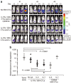
In vivo expansion of IL-7Rα+EBV-CTLs. SCID mice engrafted subcutaneously with EBV-LCLs (10 × 106 cells) were injected intravenously either with control or IL-7Rα+EBV-CTLs (10 × 106 cells/mouse). To track their homing and in vivo expansion, EBV-CTLs were transduced with a γ-retroviral vector encoding for eGFP-FFLuc. CTL localization and expansion was monitored using an in vivo imaging system. For these experiments, we used EBV-CTL lines generated from three healthy EBV-seropositive donors. Mice were then divided in three groups and received intraperitoneal injection of IL-2, IL-7, or no cytokines after CTL transfer. (a) Images of representative mice receiving IL-7Rα+EBV-CTLs. The CTL signal intensity increased by day 15–21 in mice treated either with IL-2 or IL-7 as compared to mice receiving no cytokines. (b) The bioluminescence data of all mice engrafted with EBV-LCLs and treated either with control (open symbols) or IL-7Rα+EBV-CTLs (closed symbols), and receiving no cytokines or intraperitoneal injection of IL-2 or IL-7, after CTL transfer. The graph summarizes the fold expansion in bioluminescence obtained by day 15 after CTL transfer. Control and IL-7Rα+EBV-CTLs localized at the tumor site and equally expanded in mice treated with IL-2 compared to mice receiving CTLs without cytokines. On the contrary, when mice were treated with IL-7, CTL signals increased in mice infused with IL-7Rα+EBV-CTLs but not in those infused with control EBV-CTLs (P < 0.0001).
<b>Figure 6</b>
Figure 6
In vivo antitumor effect of IL-7Rα+EBV-CTLs. To evaluate the antitumor effect, SCID mice were engrafted in the peritoneum with EBV-LCLs (3 × 10⁶ cells/mouse) labeled with FFLuc, and then treated either with control or IL-7Rα+EBV-CTLs 7–10 days later. For these experiments, we used EBV-CTL lines generated from three healthy EBV-seropositive donors. Mice were divided in three groups that received no cytokines, or IL-2, or IL-7. An additional control group received no EBV-CTLs. Tumor growth was monitored using an in vivo imaging system. (a) Tumor growth in representative mice receiving IL-7Rα+EBV-CTLs. Tumor signals progressively increased in mice that did not receive CTLs or were treated with CTLs but no cytokines. These mice were euthanized by day 20–25 after CTL infusion for excessive tumor growth. On the contrary, significant control of tumor growth was observed in mice receiving IL-7Rα+EBV-CTLs that were treated either with IL-2 or IL-7. (b) Bioluminescence signal as a measurement of tumor growth by day 14 after CTL infusions in mice receiving control or IL-7Rα+EBV-CTLs. Tumor bioluminescence rapidly increased in mice that did not receive CTLs, or received IL-7Rα+EBV-CTLs but no cytokines. On the contrary, tumor growth was controlled for at least 2 weeks when mice received IL-7Rα+EBV-CTLs and either IL-2 or IL-7 (P < 0.0001). Similar tumor control was observed in mice receiving control EBV-CTLs and IL-2, but not in mice that received control EBV-CTLs and IL-7.

References

    1. Dudley ME., and , Rosenberg SA. Adoptive-cell-transfer therapy for the treatment of patients with cancer. Nat Rev Cancer. 2003;3:666–675. - PMC - PubMed
    1. Rooney CM, Smith CA, Ng CY, Loftin SK, Sixbey JW, Gan Y, et al. Infusion of cytotoxic T cells for the prevention and treatment of Epstein-Barr virus-induced lymphoma in allogeneic transplant recipients. Blood. 1998;92:1549–1555. - PubMed
    1. Bollard CM, Aguilar L, Straathof KC, Gahn B, Huls MH, Rousseau A, et al. Cytotoxic T lymphocyte therapy for Epstein-Barr virus+ Hodgkin's disease. J Exp Med. 2004;200:1623–1633. - "V体育官网" PMC - PubMed
    1. Straathof KC, Bollard CM, Popat U, Huls MH, Lopez T, Morriss MC, et al. Treatment of nasopharyngeal carcinoma with Epstein-Barr virus--specific T lymphocytes. Blood. 2005;105:1898–1904. - "VSports" PubMed
    1. Comoli P, Pedrazzoli P, Maccario R, Basso S, Carminati O, Labirio M, et al. Cell therapy of stage IV nasopharyngeal carcinoma with autologous Epstein-Barr virus targeted cytotoxic T lymphocytes. J Clin Oncol. 2005;23:8942–8949. - PubMed

Publication types (VSports注册入口)

LinkOut - more resources