VSports在线直播 - Skip to main page content
U.S. flag

An official website of the United States government

Dot gov

The . gov means it’s official. Federal government websites often end in VSports app下载. gov or . mil. Before sharing sensitive information, make sure you’re on a federal government site. .

Https

The site is secure. The https:// ensures that you are connecting to the official website and that any information you provide is encrypted and transmitted securely V体育官网. .

. 2009 Mar 1;69(5):1792-9.
doi: 10.1158/0008-5472.CAN-08-2304. Epub 2009 Feb 17.

Overexpression of the cellular DEK protein promotes epithelial transformation in vitro and in vivo

Affiliations

Overexpression of the cellular DEK protein promotes epithelial transformation in vitro and in vivo

VSports app下载 - Trisha M Wise-Draper et al. Cancer Res. .

Abstract

High levels of expression of the human DEK gene have been correlated with numerous human malignancies. Intracellular DEK functions have been described in vitro and include DNA supercoiling, DNA replication, RNA splicing, and transcription. We have shown that DEK also suppresses cellular senescence, apoptosis, and differentiation, thus promoting cell growth and survival in monolayer and organotypic epithelial raft models VSports手机版. Such functions are likely to contribute to cancer, but direct evidence to implicate DEK as an oncogene has remained elusive. Here, we show that in line with an early role in tumorigenesis, murine papilloma formation in a classical chemical carcinogenesis model was reduced in DEK knockout mice. Additionally, human papillomavirus E6/E7, hRas, and DEK cooperated in the transformation of keratinocytes in soft agar and xenograft establishment, thus also implicating DEK in tumor promotion at later stages. Finally, adenoviral DEK depletion via short hairpin RNA expression resulted in cell death in human tumor cells in vitro and in vivo, but did not significantly affect differentiated epithelial cells. Taken together, our data uncover oncogenic DEK activities as postulated from its frequent up-regulation in human malignancies, and suggest that the targeted suppression of DEK may become a strategic approach to the treatment of cancer. .


Figures
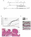
Fig. 1
Fig. 1. Dek expression is important for the formation of papillomas in vivo
A. Targeting of the mouse Dek gene. The top line shows the 5′ half of the Dek gene comprising exons 1-6. The arrow indicates the direction of transcription. The Nsi1 site (N) in exon 6 is indicated as well as the EcoRI sites (E) used for Southern blotting. The middle line shows the targeting construct containing 8.6 kb of genomic DNA with the IRES-LacZ-Neo selectable marker inserted into the NsiI site in exon 6. The bottom line shows the targeted allele. P indicates the 320 bp EcoRI-BamHI fragment used as a probe for Southern blotting. B. DMBA/TPA carcinogenesis model. Dek wildtype versus heterozygote and knockout littermates (three per group) were subjected to 100ug DMBA on the flank, and starting at one week post tumor initiation, 30ug of the tumor promoter TPA was applied twice a week for 20 weeks. The graph represents the number of papillomas per mouse that were present at the indicated time points. The asterisk represents a p value of less than 0.05 between the knockout and heterozygous mice. H&E staining below depicts the morphology of a representative papilloma at 50x magnification. The boxed area in the left panel is shown at 200x magnification in the right panel. Different genotypes developed papillomas that were morphologically similar. C. Western blot analysis. Primary keratinocytes from the heterozygous and knockout Dek mice were harvested for the isolation of protein, and western blot analysis for DEK and actin was carried out. Primary murine keratinocytes were prepared from the skin of newborn DEK heterozygous and knockout mice, floated overnight at 4°C in 0.25% trypsin/EDTA. On the next day, the epidermis was separated from the dermis, minced, filtered and keratinocytes were plated in Defined Keratinocyte Serum-Free Media supplemented with FBS and antibiotics. After seven days, protein was harvested for western blot analysis. Bands shown were imaged from the same exposed film. Immunohistochemistry on normal, untreated skin of wild type, heterozygote and knockout Dek mice confirms DEK loss in the knockout mice at 1000x magnification. Tissue sections were deparaffinized, rehydrated, subjected to antigen retrieval and processed. Sections were incubated with IgG1 antibody as a control, or with DEK antibody. Sections were washed and incubated in ABC Reagent for 30 minutes, washed again and incubated with 3,3-diaminobenzidine (DAB). Sections were then washed and stained in Nuclear Fast Red prior to mounting and visualization.
Fig. 2
Fig. 2. Overexpression of DEK protein in normal immortalized keratinocytes stimulates transformation in vitro
A. Western blot analyses. NIKs were transduced with either R780 or R780-DEK and sorted for GFP expression. Sorted NIKs were subsequently transduced with oncogenic hRas-V12 and either HPV E7 alone or HPV E6 and E7 in combination. Total protein lysates were subjected to DEK, cyclin A, p53, p21, PCNA and actin specific western blot analyses. B. Soft agar colony assay. Cells from (A) were submerged in 0.4% agarose, overlaid on top of a 0.8% agarose underlay and monitored for colony formation over three weeks, at which point colonies were counted. Two asterisks indicate a p value of less than 0.01.
Fig. 3
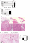
Fig. 3. DEK overexpression stimulates transformation in vivo in cooperation with the HPV oncoproteins and oncogenic ras
A, B. Nude mouse injections. 5× 10⁶ NIKs transduced with hRas, either E7 or E6/E7 and either empty R780 or R780-DEK vector were injected into the flanks of athymic nude mice. The R780 controls were injected into the left flank while DEK overexpressing cells were injected on the right flank of each mouse. Tumor formation (A) as well as tumor volume (B) were monitored over two months. Tumor volume was calculated using the following formula: length × width2 × π/6. One asterisk represents a p-value of < 0.05 while two asterisks represent a p-value of < 0.01. Experiments containing 3-4 mice per group were performed three independent times. C. Tumor morphology in the presence and absence of overexpressed DEK. Tumors from (A) were fixed, embedded in paraffin and sectioned. Sections were then stained with hematoxylin/eosin and pictures were taken at 50 and 200 fold magnification for the top and bottom panels, respectively. D. Immunohistochemistry for phosphorylated (Ser10) Histone H3 was performed on paraffin sections that were baked, deparaffinized, rehydrated and subjected to antigen retrieval. Sections were blocked using goat antiserum in phosphate buffered saline. Primary phosphorylated (Ser10) Histone H3 1:1000 (US Biological) antibodies were diluted in blocking solution, applied to tissue sections and incubated overnight at 4°C Antibody staining was detected with Vectastain Elite ABC and DAB Substrate Kits (Vector Laboratories, Inc.). Counts represent evaluation of 300 tumor cells representing 2-5 tumor sections/mouse and 3 mice per group. The asterisk represents a p value of less than 0.05, and pictures were taken at 1000 fold magnification.
Fig. 4
Fig. 4. DEK depletion in xenograft tumors results in cell death in vivo
A. In vivo imaging system (IVIS) analysis. Nude mice were injected with 5×10^5 U2OS tumor cells into each flank and injected with either empty Ad (left) or AdDEKsh (right) when tumors reached 200mm3. GFP expression in the tumors was determined and analyzed by IVIS software. Color bars represent minimum to maximum level of GFP expression. B. Immunofluorescence. Tumors from (A) were harvested, fixed and embedded in paraffin and sectioned for analysis. Sections were analyzed by immunofluorescence microscopy for the detection of cell death. The experiment was performed twice, using four mice for each experiment. Similar results were seen in both experiments.
Fig. 5
Fig. 5. Epithelial differentiation rescues cells from DEKsh-induced apoptosis
A. HFKs and U2OS cells were infected with either empty Ad or AdDEKsh and subjected to anti-active caspase 3 antibody and flow cytometry on day three. The cells were photographed after four days. B. HFK differentiation. HFKs were infected as in (A) and either subjected to normal medium, or differentiated upon the addition of 1mM calcium and 10% FBS. Cells were harvested for apoptosis assays as in A, subjected to DEK, p21, ΔNp63 and actin specific western blot analyses on day three, and analyzed for cellular morphology. One asterisk represents a p-value of < 0.05 while two asterisks represent a p-value of < 0.01. The Ad infected populations were not statistically different from each other nor were the Ad and DEKsh infected differentiated cell populations.

References

    1. von Lindern M, Fornerod M, van Baal S, et al. The translocation (6;9), associated with a specific subtype of acute myeloid leukemia, results in the fusion of two genes, dek and can, and the expression of a chimeric, leukemia-specific dek-can mRNA. Mol Cell Biol. 1992;12(4):1687–97. - PMC - PubMed
    1. Wu Q, Hoffmann MJ, Hartmann FH, Schulz WA. Amplification and overexpression of the ID4 gene at 6p22.3 in bladder cancer. Mol Cancer. 2005;4(1):16. - V体育安卓版 - PMC - PubMed
    1. Kroes RA, Jastrow A, McLone MG, et al. The identification of novel therapeutic targets for the treatment of malignant brain tumors. Cancer Lett. 2000;156(2):191–8. - VSports在线直播 - PubMed
    1. Carro MS, Spiga FM, Quarto M, et al. DEK Expression is controlled by E2F and deregulated in diverse tumor types. Cell Cycle. 2006;5(11):1202–7. - PubMed
    1. Wise-Draper TM, Allen HV, Thobe MN, et al. The human DEK proto-oncogene is a senescence inhibitor and an upregulated target of high-risk human papillomavirus E7. J Virol. 2005;79(22):14309–17. - PMC - PubMed

V体育2025版 - Publication types

"V体育安卓版" MeSH terms

Substances