Skip to main page content
U.S. flag

An official website of the United States government

Dot gov

The VSports app下载. gov means it’s official. Federal government websites often end in . gov or . mil. Before sharing sensitive information, make sure you’re on a federal government site. .

Https

The site is secure. The https:// ensures that you are connecting to the official website and that any information you provide is encrypted and transmitted securely V体育官网. .

. 2009 Apr 24;284(17):11285-92.
doi: 10.1074/jbc.M900461200. Epub 2009 Feb 16.

Regulation of neuronal cell death by MST1-FOXO1 signaling

Affiliations

"VSports注册入口" Regulation of neuronal cell death by MST1-FOXO1 signaling

Zengqiang Yuan et al. J Biol Chem. .

"V体育官网" Abstract

The protein kinase mammalian Sterile 20-like kinase 1 (MST1) plays a critical role in the regulation of cell death VSports手机版. Recent studies suggest that MST1 mediates oxidative stress-induced neuronal cell death by phosphorylating the transcription factor FOXO3 at serine 207, a site that is conserved in other FOXO family members. Here, we show that MST1-induced phosphorylation of FOXO1 at serine 212, corresponding to serine 207 in FOXO3, disrupts the association of FOXO1 with 14-3-3 proteins. Accordingly, MST1 mediates the nuclear translocation of FOXO1 in primary rat cerebellar granule neurons that are deprived of neuronal activity. We also find a requirement for MST1 in cell death of granule neurons upon withdrawal of growth factors and neuronal activity, and MST1 induces cell death in a FOXO1-dependent manner. Finally, we show that the MST1-regulatory, scaffold protein Nore1 is required for survival factor deprivation induced neuronal death. Collectively, these findings define MST1-FOXO1 signaling as an important link survival factor deprivation-induced neuronal cell death with implications for our understanding of brain development and neurological diseases. .


Figures

FIGURE 1.
FIGURE 1.
MST1 phosphorylates FOXO1 in vitro. A, lysates of 293T cells transfected with an expression plasmid encoding FLAG-MST1, kinase-dead FLAG-MST1 K59R, or the control plasmid were immunoprecipitated with the FLAG antibody and subjected to an in vitro kinase assay using full-length GST-FOXO1 as substrate in the presence of 32P-labeled ATP. Reactions were analyzed by SDS-PAGE followed by autoradiography. Auto-p-MST1 denotes autophosphorylated MST1; p-FOXO1 denotes phosphorylated FOXO1. The lower panels show the expression of FLAG-MST1 and FLAG-MST1 K59R by immunoblotting with the FLAG antibody. CMV, cytomegalovirus. B, in vitro kinase assays were carried out by incubating recombinant MST1 (rMST1) (Upstate Biotechnology) together with recombinant GST or GST-FOXO1 as substrate in the presence of 32P-labeled ATP. Reactions were analyzed by SDS-PAGE followed by autoradiography. Recombinant MST1 phosphorylated FOXO1 in vitro. C, immunoprecipitated MST1 was subjected to an in vitro kinase assay as in A using the forkhead domain of FOXO1 fused to GST (GST-FOXO1(FD)) as substrate. D, MST1 was immunoprecipitated from 293T cells and subjected to in vitro kinase assays as in A using wild type or serine 212 mutants of GST-FOXO1(FD). The designation 3A refers to the S212A, S234A, S235A FOXO1(FD) mutant; the designation 4A refers to the S212A, S218A, S234A, and S235A FOXO1(FD) mutant. Reaction products were analyzed by SDS-PAGE followed by autoradiography. Immunoblotting with the GST antibody is shown in the lower panel. Mutation of serine 212 dramatically reduced MST1-induced phosphorylation of FOXO1(FD).
FIGURE 2.
FIGURE 2.
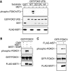
MST1 phosphorylates FOXO1 at serine 212 in vitro and in vivo. A, immunoprecipitated MST1 subjected to kinase assays using wild type or serine 212 mutants of GST-FOXO1(FD). Proteins were analyzed by immunoblotting using the phospho-FOXO (top panel), GST (middle panel), or FLAG antibody (lower panel). MST1 induced phosphorylation of FOXO1 at serine 212. The designation 4A refers to the S212A, S218A, S234A, and S235A FOXO1(FD) mutant. B, lysates of 293T cells transfected with an expression plasmid encoding FLAG-MST1, kinase-dead FLAG-MST1 K59R, or the control plasmid were immunoprecipitated with the FLAG antibody and subjected to an in vitro cold kinase assay using full-length GST-FOXO1 as substrate. Reactions were analyzed by SDS-PAGE followed by immunoblotting with the phospho-FOXO antibody. GST-FOXO1 protein levels were detected with the GST antibody. The lower panels show the expression of FLAG-MST1 and FLAG-MST1 K59R by immunoblotting with the FLAG antibody. CMV, cytomegalovirus. C, lysates of 293T cells transfected with the GFP-FOXO1 expression plasmid together with the FLAG-MST1 expression plasmid or its control vector were immunoblotted with the phospho-FOXO (top panel), GFP (middle panel), or FLAG antibody (lower panel). MST1 expression induced the phosphorylation of FOXO1 at serine 212 in cells.
FIGURE 3.
FIGURE 3.
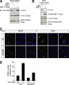
MST1-induced phosphorylation of FOXO1 disrupts FOXO1's interaction with 14-3-3 proteins and promotes the nuclear accumulation of FOXO1. A, lysates of 293T cells transfected with the GFP-FOXO1 expression plasmid together with an expression vector encoding FLAG-MST1, FLAG-MST1 K59R, or the control vector were immunoprecipitated (IP) with the GFP antibody followed by immunoblotting with a 14-3-3 antibody (top panel). The total 14-3-3 and GFP-FOXO is shown in the middle and lower panels. MST1, but not the kinase-dead MST1 K59R, disrupted FOXO1's interaction with 14-3-3 proteins. CMV, cytomegalovirus. B, lysates of 293T cells transfected with a mammalian expression vector encoding GST-FOXO1(FD) together with the MST1 expression plasmid or its control vector were subjected to GST pull-down assays followed by immunoblotting with the 14-3-3 antibody (top panel). The total MST1, 14-3-3, and FOXO1(FD) is shown in the lower panels. C, granule neurons transfected with GFP-FOXO1 together with the MST1 RNAi or control U6 plasmid maintained in membrane-depolarizing medium (30 mm KCl) or deprived of membrane depolarization (5 mm KCl) were analyzed by fluorescence microscopy. Representative images are shown (left, GFP-FOXO1; center, Hoechst; right, merged). Scale bar, 10 μm. Arrowheads indicate neurons displaying nuclear localization of FOXO1. D, quantification of results shown in Fig. 3C. MST1 knockdown significantly decreased GFP-FOXO1 nuclear accumulation in activity-deprived neurons. For each experiment, at least 100 neurons were counted (n = 3; p < 0.0001, ANOVA followed by Fisher's PLSD post hoc test).
FIGURE 4.
FIGURE 4.
MST1-FOXO1 signaling promotes cell death in neurons upon survival factor deprivation. A, granule neurons were transfected with the MST1 RNAi or control U6 plasmid together with the β-galactosidase expression vector. After 72 h, cultures were left in full survival (conditioned medium (CM)) or deprived of serum and membrane depolarization for 24 h. Transfected granule neurons were analyzed for cell survival and death based on the integrity of neuronal processes and nuclear morphology (see “Experimental Procedures”). The percentage of cell death in transfected β-galactosidase-positive-staining neurons is represented as mean ± S.E. Cell death was significantly reduced in MST1 knockdown neurons as compared with U6 control neurons upon serum and activity deprivation (n = 4; p < 0.05, ANOVA followed by Fisher's PLSD post hoc test). B, neurons transfected with FLAG-MST1 or FLAG-MST1R plasmid and the β-galactosidase expression vector were treated and analyzed as in A. MST1 encoded by the RNAi-resistant cDNA (MST1R), but not MST1 encoded by wild-type cDNA (MST1), blocked the ability of MST1 RNAi to protect neurons from serum- and activity deprivation-induced cell death (n = 3; p < 0.0001; ANOVA followed by Fisher's PLSD post hoc test). CMV, cytomegalovirus. C, neurons transfected with myr-MST1 or its control vector, together with the FOXO1, FOXO1-DN, or control expression plasmid, were analyzed as in A. FOXO1-DN, but not FOXO1, blocked the ability of myr-MST1 to promote neuronal death (n = 3; p < 0.05; ANOVA followed by Fisher's PLSD post hoc test). D–F, granule neurons were transfected with the 3×IRS (IGFBP1)-luciferase reporter gene (D), BIM-luciferase reporter gene (E), or FasL-luciferase reporter gene (F) together with the tk-Renilla reporter gene, which served as an internal control for transfection efficiency. After 24 h, cultures were left in full conditioned medium, deprived of serum and membrane depolarization (S/K-), or treated with H2O2 for 24 h. Neurons were subjected to luciferase assays. The mean ± S.E. of normalized firefly/Renilla luciferase values (RLU) relative to neurons maintained in conditioned medium is shown in the graphs (n = 3). Withdrawal of serum and membrane depolarization increased the level of 3×IRS, BIM, and FasL reporter genes (p < 0.05; ANOVA followed by Fisher's PLSD post hoc test). Hydrogen peroxide significantly stimulated the 3×IRS and BIM reporter genes (p < 0.05; ANOVA followed by Fisher's PLSD post hoc test) but failed to induce the FasL reporter gene.
FIGURE 5.
FIGURE 5.
The MST1 regulatory scaffold protein Nore1 plays distinct roles in neuronal death. A, lysates of 293T cells transfected with the FLAG-Nore1 and FLAG-MST1 expression vectors were immunoprecipitated (IP) using a rabbit polyclonal antibody to Nore1 and immunoblotted (WB) using the FLAG antibody. The total FLAG-Nore1 and FLAG-MST1 is shown in the lower panel. B, lysates of 293T cells transfected with the FLAG-Nore1 expression plasmid together with an RNAi plasmid encoding Nore1 shRNAs or the control U6 plasmid were immunoblotted with the Nore1 or ERK1/2 antibody. Nore1 RNAi triggered robust knockdown of Nore1. C, granule neurons transfected with expression plasmids encoding GFP-Nore1 and β-galactosidase (β-gal) together with the Nore1 RNAi plasmid or control U6 plasmid were subjected to immunocytochemical analysis using a mouse monoclonal antibody to β-galactosidase and a rabbit polyclonal antibody to GFP. Nore1 RNAi induced the knockdown of Nore1 in nearly all cells examined. D, granule neurons were transfected with the Nore1 RNAi plasmid or control U6 plasmid together with a plasmid encoding β-galactosidase. After 72 h, cultures were left untreated or treated with H2O2, and 24 h later cultures were analyzed as described in Fig. 4A. Nore1 knockdown sensitized neurons to H2O2-induced cell death as compared with control (n = 3, p < 0.05, ANOVA followed by Fisher's PLSD post hoc test). CM, conditioned medium. E, granule neurons were transfected with the Nore1 RNAi plasmid or control U6 plasmid together with the β-galactosidase expression plasmid. After 72 h, cultures were deprived of survival factors and left untreated for 24 h and analyzed as in Fig. 4A. Nore1 knockdown protected neurons from survival factor deprivation-induced cell death as compared with control (n = 3, p < 0.05, t test).

References

    1. Oppenheim, R. (1989) Annu. Rev. Neurosci. 14, 453-501 - PubMed
    1. Pettmann, B., and Henderson, C. E. (1998) Neuron 20, 633-647 - PubMed
    1. Yuan, J., and Yankner, B. A. (2000) Nature 407, 802-809 - PubMed
    1. Mattson, M. P. (2000) Nat. Rev. Mol. Cell Biol. 1, 120-129 - V体育安卓版 - PubMed
    1. Choi, D. W. (1996) Curr. Opin. Neurobiol. 6, 667-672 - PubMed

Publication types

MeSH terms