Skip to main page content
U.S. flag

An official website of the United States government

Dot gov

The . gov means it’s official. Federal government websites often end in VSports app下载. gov or . mil. Before sharing sensitive information, make sure you’re on a federal government site. .

Https

The site is secure V体育官网. The https:// ensures that you are connecting to the official website and that any information you provide is encrypted and transmitted securely. .

. 2009 Jun;5(2):129-37.
doi: 10.1007/s11302-009-9141-7. Epub 2009 Feb 12.

The P2X(7) receptor-pannexin connection to dye uptake and IL-1beta release

Affiliations

The P2X(7) receptor-pannexin connection to dye uptake and IL-1beta release

Pablo Pelegrin (V体育官网) et al. Purinergic Signal. 2009 Jun.

Abstract

The P2X(7) receptor (P2X(7)R) is uniquely associated with two distinct cellular responses: activation of a dye-permeable pathway allowing passage of molecules up to 900 Da and rapid release of the pro-inflammatory cytokine, interleukin-1beta (IL-1beta), from activated macrophage VSports手机版. How this dye uptake path forms and whether it is involved in IL-1beta release has not been known. Pannexin-1 is a recently identified protein found to physically associate with the P2X(7)R. Inhibition of pannexin-1 does not alter P2X(7)R ion channel activation or associated calcium flux but blocks one component of P2X(7)R-induced dye uptake and unmasks a slower, previously undetected, dye uptake pathway. Inhibition of pannexin-1 blocks P2X(7)R-mediated IL-1beta release from macrophage as well as release mediated by other stimuli which couple to activation of capase-1 and additionally inhibits the release of interleukin-1alpha, a member of the IL-1 family whose processing does not require caspase-1 activation. Thus, pannexin-1 is linked to both dye uptake and IL-1beta release but via distinct mechanisms. .


Figures
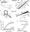
Fig. 1
Fig. 1
Evidence for and against the P2X7R ion channel itself dilating to form the dye uptake large pore. a Example of membrane current recorded in response to P2X7R stimulation in extracellular solution containing the large cation, NMDG+. Initial current is outward because NMDG+ initially is impermeable and only intracellular sodium passes outward but with time current turns inward due to NMDG+ entering the cell. b Example of reversal potential measurement experiments carried out at 2-s intervals during application of BzATP as shown in (a); reversal potential shifts to positive values which shows that NMDG+ is entering the cell. c, d Examples of the same experiment but from cell expressing mutated P2X7R; now there is no NMDG+ shift indicating no entry of NMDG+ into the cell, but the same mutated receptor shows enhanced dye uptake (e). Recording in (a) is from Virginio et al. [35] while results shown in (b)–(e) are from Jiang et al. [39]
Fig. 2
Fig. 2
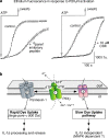
Two phases to P2X7R-mediated dye uptake revealed by inhibition of panx1. a Original traces of typical dye uptake experiments carried out on HEK 293 cells expressing rat P2X7R; each trace shows average ± SEM from 20 to 30 cells; control fluorescence (in arbitrary fluorescence units) saturates the optical system in both examples. Inhibition of panx1 with CBX or 10panx1-mimetic inhibitory peptide dramatically delays dye uptake; results detailed in [40, 42] suggest two independent processes. b Distinct roles and underlying mechanisms for the two dye uptake processes after P2X7R activation. Panx1 is involved in the initial rapid dye uptake and in IL-1β processing and release. Mechanisms underlying the slow dye uptake and its physiological significance are unknown but results from [43] suggest it may involve MAPK-dependent pathways. P2X7R cartoon is represented as a homotrimer based on studies of the mammalian P2X1 and P2X2 receptors [71]. Panx1 cartoon is represented as a plasma membrane hemichannel consisting of a complex of six subunits, hypothetically arranged based on analogy to connexin gap junction hemichannels
Fig. 3
Fig. 3
Schematic depicting distinct models of the activation of caspase-1 and IL-1β release induced by P2X7R and panx1 activation. Toll-like receptor (TLR) activation by bacterial PAMPs activates the production of pro-IL-1β as well as enhances the expression and activity of P2X7R and panx1. Under resting conditions, P2X7R may keep panx1 under negative regulation via direct interaction. High concentrations of extracellular ATP activates P2X7R ion channel resulting in a cation permeability (Na+ influx/K+ efflux and intracellular Ca++ rise) and the activation of panx1. Panx1 activation allows a rapid dye uptake permeabilization pathway and potentially the entry of either bacterial PAMPs and/or extracellular ATP that will directly activate NLRP3 (nucleotide-binding domain and leucine-rich repeat containing a pyrin domain) molecule using the adaptor molecule ASC (apoptosis-associated speck-like protein containing a CARD). Activated NLRP3 with ASC aggregates pro-caspase-1 molecules through the formation of a macromolecular inflammasome complex that facilitates subsequent pro-caspase-1 activation and pro-IL-1β processing to its bioactive state. NLRP3 inflammasome complex cartoon is based on studies on the NLRP1 inflammasome and is probably comprised of five to seven subunits, each subunit consisting of NLRP3 and ASC with the ability to recruit five to seven pro-caspase-1 molecules [68]. Inflammasome pictorial adapted from Ting et al. [72]

References

    1. {'text': '', 'ref_index': 1, 'ids': [{'type': 'DOI', 'value': '10.1016/0006-291X(75)90207-7', 'is_inner': False, 'url': '"V体育2025版" https://doi.org/10.1016/0006-291x(75)90207-7'}, {'type': 'PubMed', 'value': '1039', 'is_inner': True, 'url': 'https://pubmed.ncbi.nlm.nih.gov/1039/'}]}
    2. Rozengurt E, Heppel LA (1975) A specific effect of ATP on the permeability of transformed 3T3 cells. Biochem Biophys Res Commun 67:1581–1588 - PubMed (V体育平台登录)
    1. {'text': '', 'ref_index': 1, 'ids': [{'type': 'DOI', 'value': '10.1038/279541a0', 'is_inner': False, 'url': 'https://doi.org/10.1038/279541a0'}, {'type': 'PubMed', 'value': '450099', 'is_inner': True, 'url': 'https://pubmed.ncbi.nlm.nih.gov/450099/'}]}
    2. Cockcroft S, Gomperts BD (1979) ATP induces nucleotide permeability in rat mast cells. Nature 279:541–542 - PubMed
    1. {'text': '', 'ref_index': 1, 'ids': [{'type': 'DOI', 'value': '10.1073/pnas.85.21.7988', 'is_inner': False, 'url': 'https://doi.org/10.1073/pnas.85.21.7988'}, {'type': 'PMC', 'value': 'PMC282339', 'is_inner': False, 'url': 'https://pmc.ncbi.nlm.nih.gov/articles/PMC282339/'}, {'type': 'PubMed', 'value': '3186701', 'is_inner': True, 'url': 'https://pubmed.ncbi.nlm.nih.gov/3186701/'}]}
    2. Buisman HP, Steinberg TH, Fischbarg J, Silverstein SC, Vogelzang SA, Ince C, Ypey DL, Leijh PCJ (1988) Extracellular ATP induces a large nonselective conductance in macrophage plasma membranes. Proc Natl Acad Sci U S A 85:7988–7992 - PMC - PubMed
    1. {'text': '', 'ref_index': 1, 'ids': [{'type': 'PubMed', 'value': '2932438', 'is_inner': True, 'url': 'https://pubmed.ncbi.nlm.nih.gov/2932438/' (VSports手机版)}]}
    2. Sung SS, Young JD, Origlio AM, Heiple JM, Kaback HR, Silverstein SC (1985) Extracellular ATP perturbs transmembrane ion fluxes, elevates cytosolic [Ca2+] and inhibits phagocytosis in mouse macrophages. J Biol Chem 260:13442–13449 - PubMed
    1. {'text': '', 'ref_index': 1, 'ids': [{'type': 'PubMed', 'value': '2950094', 'is_inner': True, 'url': 'https://pubmed.ncbi.nlm.nih.gov/2950094/'}]}
    2. Steinberg TH, Silverstein SC (1987) Extracellular ATP4− promotes cation fluxes in the J774 mouse macrophage cell line. J Biol Chem 262:3118–3122 - PubMed

LinkOut - more resources