Skip to main page content (V体育2025版)
U.S. flag

An official website of the United States government

Dot gov

The . gov means it’s official. Federal government websites often end in . gov or . mil. Before sharing sensitive information, make sure you’re on a federal government site. VSports app下载.

Https

The site is secure V体育官网. The https:// ensures that you are connecting to the official website and that any information you provide is encrypted and transmitted securely. .

. 2009 Feb;5(2):e1000288.
doi: 10.1371/journal.ppat.1000288. Epub 2009 Feb 6.

Disruption of the Toxoplasma gondii parasitophorous vacuole by IFNgamma-inducible immunity-related GTPases (IRG proteins) triggers necrotic cell death

Affiliations

Disruption of the Toxoplasma gondii parasitophorous vacuole by IFNgamma-inducible immunity-related GTPases (IRG proteins) triggers necrotic cell death

Yang O Zhao et al. PLoS Pathog. 2009 Feb.

V体育官网入口 - Abstract

Toxoplasma gondii is a natural intracellular protozoal pathogen of mice and other small mammals VSports手机版. After infection, the parasite replicates freely in many cell types (tachyzoite stage) before undergoing a phase transition and encysting in brain and muscle (bradyzoite stage). In the mouse, early immune resistance to the tachyzoite stage is mediated by the family of interferon-inducible immunity-related GTPases (IRG proteins), but little is known of the nature of this resistance. We reported earlier that IRG proteins accumulate on intracellular vacuoles containing the pathogen, and that the vacuolar membrane subsequently ruptures. In this report, live-cell imaging microscopy has been used to follow this process and its consequences in real time. We show that the rupture of the vacuole is inevitably followed by death of the intracellular parasite, shown by its permeability to cytosolic protein markers. Death of the parasite is followed by the death of the infected cell. The death of the cell has features of pyronecrosis, including membrane permeabilisation and release of the inflammatory protein, HMGB1, but caspase-1 cleavage is not detected. This sequence of events occurs on a large scale only following infection of IFNgamma-induced cells with an avirulent strain of T. gondii, and is reduced by expression of a dominant negative mutant IRG protein. Cells infected by virulent strains rarely undergo necrosis. We did not find autophagy to play any role in the key steps leading to the death of the parasite. We conclude that IRG proteins resist infection by avirulent T. gondii by a novel mechanism involving disruption of the vacuolar membrane, which in turn ultimately leads to the necrotic death of the infected cell. .


Conflict of interest statement

The authors have declared that no competing interests exist.

Figures

Figure 1
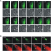
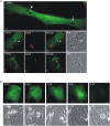
Figure 1. Disruption of IRG-positive T. gondii vacuoles.
MEFs were transfected with an expression plasmid encoding Irga6-ctag1-EGFP and treated with 200 U/ml IFNγ. 24 hours later cells were infected with ME49 strain T. gondii at an MOI between 5 and 10 for 1 hour and observed microscopically by time-lapse photography. Images were taken at 1 minute (A and B) or 3 minute (C) intervals. In C, both fluorescence and corresponding phase contrast images are shown. Selected images from the series show the disruption of Irga6-ctag1-EGFP positive T. gondii vacuoles. The number of PVs in individual cells were 3, 4 and 2 for A, B and C, respectively. Disruption of IRG-positive PVM is indicated by arrows. It was frequently observed that vacuoles and the included parasites tended to round up shortly before disruption (A and C: arrowheads). The timing is indicated as hour:minute post infection. Scale bar: 5 µm. The videos from which these frames were extracted are presented as Video S1(Fig. 1A), S2(Fig. 1B), and S5 and S6(Fig. 1C).
Figure 2
Figure 2. T. gondii is permeabilised in the host cytosol after the disruption of PVM.
(A) MEFs were induced with 200 U/ml IFNγ and transfected with pEGFP. 24 hours later they were infected with ME49 strain T. gondii at an MOI between 5 and 10 for 2 hours and observed microscopically by time-lapse photography. One intracellular parasite was observed throughout the experiment. Images were taken at 5 minute intervals, and selected images from the series (EGFP and phase contrast) are shown. The intracellular T. gondii, initially seen as a dark form excluding EGFP, became permeable to EGFP between 3:45 and 3:55 after infection (arrowheads). At the same time there was a detectable change in the appearance of the parasite in the phase contrast series from dense to light at 3:25 that probably corresponded to the rupture of the vacuole. Arrows indicate the rounding up of T. gondii also noted in Fig. 1A and C. (B) MEFs were treated with IFNγ and co-transfected with Irga6-ctag1-EGFP and pCherry for 24 hours. Cells were then infected with ME49 strain T. gondii for 1 hour and observed by time-lapse microscopy. Two PVs were observed within this cell. Images were taken at 3 minute intervals, and selected images from the series are shown (upper panel Irga6-ctag1-EGFP and lower panel Cherry). The T. gondii PVM was disrupted at 1:39 (arrows) and the parasite was permeabilised at 2:06 (arrowheads). These images partially overlap with and extend the series shown in Fig. 1C. Scale bar: 5 µm. The videos from which these frames were extracted are presented as Videos S3 and S4(Fig. 2A) and S5, S6, S7, and S8(Fig. 2B).
Figure 3
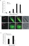
Figure 3. Irgb6 contributes to the IFNγ-dependent killing of T. gondii.
(A) MEFs were treated with 200 U/ml IFNγ (black bar) or left untreated (white bar) and transfected with pEGFP. After 24 hours cells were infected with ME49 strain T. gondii at MOI between 5 and 10 for the indicated times, fixed and stained for GRA7 to identify intracellular T. gondii. Permeabilised T. gondii containing EGFP were counted and are shown as the percentage of the total number of intracellular parasites counted in EGFP-transfected cells at each time point. Means and ranges of values from two experiments are shown. (B) MEFs were treated with 200 U/ml IFNγ and co-transfected with pEGFP and expression plasmids encoding Irgb6-wt-FLAG (a–d) or Irgb6-K69A-FLAG (e–h). 4 hours after infection with ME49 strain T. gondii at an MOI between 5 and 10, cells were fixed and stained for FLAG tag (red). Note the typical cytoplasmic aggregates of Irgb6-K69A-FLAG not associated with the T. gondii PV, and the very weak staining of the PVM itself, as documented elsewhere . Arrows indicate a permeabilised T. gondii in a disrupted vacuole while the arrowheads indicate EGFP-impermeable parasites in intact vacuoles. Scale bar 10 µm. (C) Quantification of permeabilised T. gondii at 2 and 4 hours after infection as described in (B). Means and ranges of values from two experiments are shown. 100–200 PVs per data point were counted blind.
Figure 4
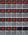
Figure 4. Host cells die after disruption of the PVM and death of the T. gondii.
MEFs were treated with 200 U/ml IFNγ and co-transfected with Irga6-ctag1-EGFP and pDsRed constructs for 24 hours. Cells were then infected with ME49 strain T. gondii at MOI between 5 and 10 for 1 hour and observed microscopically by time-lapse photography. Three PVs were observed within this cell when the experiment started. Images were taken at 5 minutes intervals, and the complete series is shown. Two Irga6-ctag1-EGFP-positive PVMs were disrupted at 1:10 and 1:40, respectively (arrows). These two T. gondii became permeable to mDsRed at 1:30 and 2:25, respectively (arrowheads). At 2:30 after infection, the mDsRed signal suddenly disappeared from the host cell accompanied by a drastic change in host cell morphology shown in phase contrast images. Scale bar: 10 µm. The videos from which these frames were extracted are presented as Videos S9 and S10.
Figure 5
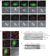
Figure 5. IFNγ-treated T. gondii infected host cells undergo necrosis after vacuolar disruption.
(A) Phosphatidylserine is not expressed on the cell surface before death. MEFs were induced with 200 U/ml IFNγ and transfected with pEGFP. After 24 hours they were infected with ME49 strain T. gondii for 1 hour at MOI between 5 and 10 and observed microscopically by time-lapse photography. Phosphatidylserine was detected by adding 1% (v/v) Alexa-555-labeled annexin V with 2.5 mM CaCl2 into the medium. Images were taken at 5 minute intervals and selected images from the series are shown. One intracellular T. gondii rounded up and was permeabilised at 1:30 (arrows) and the host cell died at 2:00. No annexin V signal was detected on the plasma membrane before cell death. After permeabilisation of the plasma membrane, annexin V accumulated steadily on intracellular material. Scale bar: 10 µm. The videos from which these frames were extracted are presented as Videos S11 and S12. (B) Cytochrome C is not released from mitochondria in cells containing a disrupted vacuole. MEFs were induced with 200 U/ml IFNγ and transfected with pEGFP for 24 hours (c–f). Cells were then infected with ME49 strain T. gondii at MOI between 5 and 10 for 4 hours, fixed and stained for cytochrome C (red) and Irgb6 (inbox in d). Arrows indicate the permeabilised T. gondii and Irgb6 staining shows the disrupted PVM. As control, MEFs without any treatment (a), or treated with TNFα (40 ng/ml) and cycloheximide (10 µg/ml) for 4 hours to induce apoptosis (b), were stained for cytochrome C. Arrowheads indicate cells showing release of mitochondrial cytochrome C. Scale bar 10 µm. (C) HMGB1 is released from IFNγ-induced cells infected with T. gondii. MEFs were treated with 200 U/ml IFNγ or left untreated for 24 hours and infected with ME49 T. gondii for the indicated times. Cells were then lysed and the lysates blotted for cleaved caspase-3 and PARP, as well as calnexin as loading control. Cell culture supernatants were collected at the indicated times after infection and blotted for HMGB1. MEFs treated with TNFα (40 ng/ml) and cycloheximide (10 µg/ml) were blotted as control for apoptotic cells. (D) Caspase-1 and IL-1β are not activated. BMMs were treated as described in (C). Cell lysates were blotted for caspase-1 and cell culture supernatant were blotted for IL-1β. BMMs were first treated with LPS (1 µg/µl) for 24 hours, and then treated with nigericin (20 µM) for the indicated times to activate inflammasomes as positive control.
Figure 6
Figure 6. Macroautophagy is not involved in the disruption of the T. gondii PVM or the death of the parasite.
(A) MEFs were left uninduced (a) or induced with 200 U/ml IFNγ (b–i) and transfected with pEGFP-LC3. After 24 hours cells were infected with ME49 strain T. gondii at an MOI between 5 and 10 for 6 hours. Cells were then fixed and stained for Irga6 (red, b–i). Arrows indicate the Irga6-positive PVM and arrowheads indicate EGFP-LC3-associated T. gondii vacuoles. Scale bar 10 µm. (B) MEFs were induced with 200 U/ml IFNγ and transfected with pEGFP-LC3. After 24 hours cells were infected with ME49 strain T. gondii at an MOI between 5 and 10 for 2 hour and observed microscopically by time-lapse photography. Two PVs were observed within this cell. Images were taken at 5 minutes intervals, and selected images from the series are shown. One T. gondii was obviously permeabilised and incorporated free cytosolic EGFP-LC3 at 3:00 after infection and the host cell died at 4:35 after infection. There is no clear association between EGFP-LC3 and T. gondii vacuoles. Scale bar 10 µm. The videos from which these frames were extracted are presented as Videos S13 and S14.
Figure 7
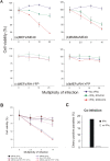
Figure 7. Virulent T. gondii are resistant to the IRG- and IFNγ-dependent cell-necrotic programme.
(A) MEFs (a, c–d) or BMMs (b) were seeded into 96 well plates and induced with the indicated dose of IFNγ for 24 hours. Cells were then infected with T. gondii avirulent strain ME49 (a–b) or virulent strain RH-YFP (c–d) for 8 hours at the indicated MOIs. Cell viabilities were measured and expressed as percentages of those recorded for uninfected cells (MOI = 0). (B) MEFs were treated as described in (A) and infected for 8 hr with ME49 or RH-YFP alone or co-infected with ME49 and RH-YFP simultaneously. Cell viabilities were measured and expressed as percentages of those recorded for uninfected cells. (C) MEFs were transfected with pCherry and induced with 200 U/ml IFNγ for 24 hours (black bar) or left untreated (white bar). Cells were then co-infected with ME49 and RH-YFP T. gondii strains (MOI 5 for each strain) for 4 hours. Permeabilised parasites containing Cherry were counted, in cells containing at least one RH-YFP parasite, as a proportion of all intracellular ME49. 200–300 PVs per data point were counted blind.
Figure 8
Figure 8. The time-course of IRG-mediated ME49 T. gondii resistance.
The figure illustrates the “typical” time course for the IRG-mediated programme of resistance against T. gondii strain ME49 in IFNγ-induced cells. The values for each step in the programme are the mean values taken from Table S1. It is important to stress that most of these timings are very variable, including the time after adding the T gondii to culture at which cell is finally infected. Only the interval between vacuole disruption and T. gondii permeabilisation is rather well defined. Nevertheless the data give a correct account of the invariable order of events and their approximate relative timing.

References (VSports app下载)

    1. Saeij JP, Boyle JP, Boothroyd JC. Differences among the three major strains of Toxoplasma gondii and their specific interactions with the infected host. Trends Parasitol. 2005;21:476–481. - PubMed
    1. Sibley LD, Boothroyd JC. Virulent strains of Toxoplasma gondii comprise a single clonal lineage. Nature. 1992;359:82–85. - VSports手机版 - PubMed
    1. Frenkel JK. Pathophysiology of toxoplasmosis. Parasitol Today. 1988;4:273–278. - PubMed (VSports在线直播)
    1. Hunter CA, Remington JS. Immunopathogenesis of toxoplasmic encephalitis. J Infect Dis. 1994;170:1057–1067. - "V体育2025版" PubMed
    1. Yap GS, Sher A. Cell-mediated immunity to Toxoplasma gondii: initiation, regulation and effector function. Immunobiology. 1999;201:240–247. - VSports最新版本 - PubMed

"V体育2025版" Publication types

"V体育平台登录" MeSH terms

Substances