<var date-time="8eriII"></var>
Skip to main page content
U.S. flag

An official website of the United States government

Dot gov

The . gov means it’s official VSports app下载. Federal government websites often end in . gov or . mil. Before sharing sensitive information, make sure you’re on a federal government site. .

Https

The site is secure V体育官网. The https:// ensures that you are connecting to the official website and that any information you provide is encrypted and transmitted securely. .

. 2009 Apr 17;284(16):10473-9.
doi: 10.1074/jbc.M809106200. Epub 2009 Feb 5.

Structural and functional analysis of a C3b-specific antibody that selectively inhibits the alternative pathway of complement

Affiliations

Structural and functional analysis of a C3b-specific antibody that selectively inhibits the alternative pathway of complement

Kenneth J Katschke Jr et al. J Biol Chem. .

V体育平台登录 - Abstract

Amplification of the complement cascade through the alternative pathway can lead to excessive inflammation. Targeting C3b, a component central to the alternative pathway of complement, provides a powerful approach to inhibit complement-mediated immune responses and tissue injury. In the present study, phage display technology was employed to generate an antibody that selectively recognizes C3b but not the non-activated molecule C3 VSports手机版. The crystal structure of C3b in complex with a Fab fragment of this antibody (S77) illustrates the structural basis for this selectivity. Cleavage of C3 to C3b results in a plethora of structural changes within C3, including the rearrangement of macroglobulin domain 6 enabling binding of S77 to the adjacent macroglobulin domain 7 domain. S77 blocks binding of factor B to C3b inhibiting the first step in the formation of the alternative pathway C3 convertase. In addition, S77 inhibits C5 binding to C3b. This results in significantly reduced formations of anaphylatoxins and membrane-attack complexes. This study for the first time demonstrates the structural basis for complement inhibition by a C3b-selective antibody and provides insights into the molecular mechanisms of alternative pathway complement activation. .


"VSports在线直播" Figures
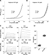
FIGURE 1.
FIGURE 1.
Generation of a phage antibody that selectively binds to C3b, but not native C3. Binding of S77 IgG (A) or a polyclonal anti-C3 antibody (C3 pAb, B) to C3b or C3 protein. Binding is expressed as the ratio of optical densities of the reaction mixtures measured at 450 and 630 nm. C, Biacore analysis of binding affinity of S77 to C3b, iC3, iC3b, and C3c. S77 was captured on a CM5 sensor chip. Increasing concentrations of S77 (3.1, 6.25, 12.5, 25, 50, and 100 nm) were injected for 180 s. The KD is calculated from binding curves showing response at equilibrium plotted against the concentration. D, the plasma concentration of C3 protein recognized by S77 IgG is significantly lower than the plasma concentration of total C3 proteins. Data in A and B represent average ± S.D. of triplicate measurements.
FIGURE 2.
FIGURE 2.
Crystal structure of S77 in complex with C3b. A, structure of the C3b-S77 dimer in the asymmetric unit. One C3b-S77 complex is shown as a ribbon diagram; the other is in surface representation. The β-chains of C3b are depicted in green, the α-chains shown in violet, light blue (TED domain), and orange (CUB domain). S77 is shown with the light chains colored white, and the heavy chains colored yellow. B, the Fab surface is shown with the light chain atoms in white and heavy chain in yellow. All amino acids that have at least one atom closer than 4.5 Å to the S77 are shown in stick representation and are labeled with carbon atoms of residues from the β-chain and α-chain colored green and violet, respectively. C, the S77 binding site of C3b. Atoms of C3b that are closer than 4.5, 4, and 3.5 Å are colored yellow, orange, and red, respectively. D, the surface of C3 is shown after superimposing the MG7 domain of C3 onto the MG7 domain of the C3b-S77 complex. Although the MG7 domains of C3 and C3b superimpose very well (r.m.s.d. 0.5 Å for 97 common Cα positions), the different relative orientation of MG7 in respect to MG6 leads to steric clashes between the light chain of the S77 and the MG6 domain in a potential complex, thus preventing S77 binding to C3.
FIGURE 3.
FIGURE 3.
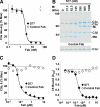
S77 inhibits fH and sCR1 binding to C3 and inhibits fH and sCR1 co-factor activity. A, microtiter plates were coated with C3b, and fH was added in the presence of increasing concentrations of S77 or control Fab. Binding of fH to C3b was determined using an anti-fH antibody and a secondary HRPO-conjugated antibody. Absorbance of the reaction mixture was measured at 450 nm. B, cofactor activity for fI-mediated cleavage of C3b was measured by incubating C3b and fI with fH or increasing concentrations of S77 or control Fab. The mixture was incubated at 37 °C, and the samples were analyzed by gel-electrophoresis and Simply Blue staining. C, S77 inhibits CR1 binding to C3b. Microtiter plates coated with C3b were incubated with sCR1 and increasing concentrations of S77 or control Fab. Binding of sCR1 to C3b was detected with an anti-CR1 primary antibody and an HRPO-conjugated secondary antibody. D, sCR1 cofactor activity was determined as described under B except that fH was replaced by sCR1. Data in A and C are expressed as mean ± S.D. of four repeats.
FIGURE 4.
FIGURE 4.
S77 affects the formation, but not stability, of the alternative pathway C3 convertase. A, S77 inhibits binding of factor B to C3b. C3b was coated on microtiter plates. S77 or control Fab was added, followed by addition of factor B. Binding of factor B to C3b was determined with anti-factor B antibody and secondary antibodies conjugated to HRPO. B, S77 Fab inhibits formation of C3 convertase. C3b was coated on microtiter plates. S77 or control Fab was added, followed by addition of fB and factor D. Binding of fBb to C3b was determined with anti-fB antibody and secondary antibody conjugated to HRPO. C, S77 does not decay the C3 convertase. Microtiter plates were coated with C3b, incubated with factor B and factor D followed by the addition of S77, sCR1 or control Fab. Factor Bb was detected with goat anti-human factor B and donkey anti-goat antibody conjugated to HRPO. Absorbance of the reaction product was read at 450 nm. Data are expressed as mean ± S.D. of four repeats.
FIGURE 5.
FIGURE 5.
S77 inhibits the alternative pathway C3 and C5 convertase. A, C3 and factors B and D were incubated in the presence of increasing concentrations of S77 or control Fab. The concentration of C3a des-Arg reaction product was determined by ELISA. Data are expressed as mean ± S.D. of triplicate measurements. B, in a separate experiment, the effect of S77 or control Fab on C3 convertase activity was determined by gel electrophoresis. Experiments were repeated three times with similar results. C, S77 inhibits C5 convertase activity. C5 convertase was assembled on the surface of zymosan particles. A fixed concentration of C5 and convertase was mixed with increasing concentrations of S77 or control Fab. Convertase activity was determined using a chicken erythrocyte hemolytic assay and expressed as percentage of hemolysis in the absence of inhibitor. D, C3b was coated on microtiter plates and incubated with a mixture of C5 and increasing concentrations of S77 or control Fab. C5 bound to plate-coated C3b was detected with a polyclonal antibody against C5 and a HRPO-conjugated secondary antibody.
FIGURE 6.
FIGURE 6.
S77 inhibits the alternative, but not classical, pathway C5 convertase in human serum. A, S77 inhibits AP-mediated hemolysis. Rabbit erythrocytes were incubated with C1q-depleted human serum in the presence of increasing concentrations of S77 or control Fab. Hemolysis was determined by absorbance of the supernatant at 412 nm. Convertase activation was expressed as percentage of hemolysis in the absence of inhibitor. B, S77 does not inhibit CP convertase activation. IgM-coated sheep erythrocytes were incubated with factor B-depleted human serum and increasing concentrations of S77 or anti-C5 antibody. C, S77 lacks complement inhibitory activity in rhesus serum where His-897 is substituted by Phe-897. CRIg-Fc was used as a positive control. D, His-897 of C3b is within hydrogen bonding distance to the main-chain atoms of the heavy chain CDR1 and CDR3 of S77. The exchange of His-897 against phenylalanine, the respective amino acid in the C3 sequence of rhesus, leads to the loss of these hydrophilic interactions. Data in A–C are expressed as mean ± S.D. of triplicate measurements.
FIGURE 7.
FIGURE 7.
Structural basis for complement inhibition by S77, CRIg, and compstatin. The superposition of the C3b-S77 complex, the C3c-CRIg complex, and the C3c-compstatin complex shows that all three inhibitors recognize the same “face” of the complement protein; their binding sites are located on the opposite site of the TED position in C3b. C3b-C3c and S77 are colored as described in Fig. 2, CRIg is shown in red, and compstatin is depicted as purple sticks.

"VSports手机版" References

    1. Thurman, J. M., and Holers, V. M. (2006) J. Immunol. 176 1305–1310 - PubMed
    1. Ricklin, D., and Lambris, J. D. (2007) Nat. Biotechnol. 25 1265–1275 - PMC - PubMed
    1. Muller-Eberhard, H. J., and Gotze, O. (1972) J. Exp. Med. 135 1003–1008 - PMC - PubMed
    1. Janssen, B. J., Christodoulidou, A., McCarthy, A., Lambris, J. D., and Gros, P. (2006) Nature 444 213–216 - PubMed
    1. Janssen, B. J., Huizinga, E. G., Raaijmakers, H. C., Roos, A., Daha, M. R., Nilsson-Ekdahl, K., Nilsson, B., and Gros, P. (2005) Nature 437 505–511 - PubMed

Publication types

MeSH terms