Skip to main page content
U.S. flag

An official website of the United States government

Dot gov

The . gov means it’s official VSports app下载. Federal government websites often end in . gov or . mil. Before sharing sensitive information, make sure you’re on a federal government site. .

Https

The site is secure V体育官网. The https:// ensures that you are connecting to the official website and that any information you provide is encrypted and transmitted securely. .

. 2009 Mar;10(3):306-13.
doi: 10.1038/ni.1696. Epub 2009 Jan 25.

VSports最新版本 - The gene encoding early growth response 2, a target of the transcription factor NFAT, is required for the development and maturation of natural killer T cells

Affiliations

The gene encoding early growth response 2, a target of the transcription factor NFAT, is required for the development and maturation of natural killer T cells

Vanja Lazarevic (V体育官网) et al. Nat Immunol. 2009 Mar.

Abstract

The influence of signals transmitted by the phosphatase calcineurin and the transcription factor NFAT on the development and function of natural killer T (NKT) cells is unclear. In this report, we demonstrate that the transcription factor early growth response 2 (Egr2), a target gene of NFAT, was specifically required for the ontogeny of NKT cells but not that of conventional CD4(+) or CD8(+) T cells VSports手机版. NKT cells developed normally in the absence of Egr1 or Egr3, which suggests that Egr2 is a specific regulator of NKT cell differentiation. We found that Egr2 was important in the selection, survival and maturation of NKT cells. Our findings emphasize the importance of the calcineurin-NFAT-Egr2 pathway in the development of the NKT lymphocyte lineage. .


VSports在线直播 - Conflict of interest statement

Competing Interests Statement: The authors declare competing financial interests: details accompany the full-text HTML version of the paper at http://www V体育安卓版. nature. com/natureimmunology/.

Figures

Figure 1
Figure 1
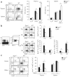
The calcineurin-NFAT pathway is required for the development of NKT cells. (a) Quantitative real-time PCR analysis of NFATc1, NFATc2 and NFATc3 mRNA transcripts in various cell populations from wild-type mice. Data are representative of two independent experiments. (b) Flow cytometry of thymocytes and splenocytes from Cnb1fl/fl Lck-Cre+ mice and control Cnb1fl/+ Lck-Cre+ littermates, stained with PBS57-loaded CD1d tetramers (tet) and anti-B220 (left), and percent tetramer-binding (Tet+) cells in the lymphocyte gate and number of live tetramer-binding cells (right). Numbers in plots indicate percent tetramer-positive B220 cells (outlined areas, left). *, P < 0.05, and **, P < 0.001 (Mann-Whitney test). Data are representative of two independent experiments with three to four mice per group per experiment (mean and s.e.m.). (c) Real-time PCR analysis of mRNA transcripts encoding Egr1, Egr2 and Egr3 in thymocytes and splenocytes stimulated for 0–6 h (horizontal axes) with PMA and ionomycin. Data are representative of two independent experiments.
Figure 2
Figure 2
Egr2−/− mice have a severe defect in NKT cell development. (a) Real-time PCR analysis of mRNA transcripts encoding Egr1, Egr2 and Egr3 in sorted NKT cells stimulated for 0–6 h (horizontal axes) with PMA and ionomycin. Data are representative of two independent experiments. (b,c) Flow cytometry of thymocytes, splenocytes and liver mononuclear cells from Egr1−/−, Egr3−/− and wild-type (WT) mice, and from Egr2−/− and wild-type fetal liver (FL) chimeras, stained with tetramers and anti-CD8. (b) Numbers in plots indicate percent tetramer-positive cells in the lymphocyte gate (outlined). (c) *, P < 0.0001 (Student's t-test). Data are representative of two to six independent experiments (mean and s.e.m. in c). (d) Real-time PCR analysis of Vα14Jα18 mRNA transcripts in thymuses from Egr2−/− and wild-type fetal liver chimeras, normalized to the quantity of transcripts encoding the constant α-chain. *, P < 0.001 (Mann-Whitney test). Data are representative of two experiments (mean and s.e.m.). (e) Real-time PCR analysis of EGR2 mRNA expression in human BM2a.3 cells stimulated for 0–6 h (horizontal axis) with PMA and ionomycin. Data are representative of one experiment (pooled results of ten mice).
Figure 3
Figure 3
Egr2 is required for the productive selection and terminal maturation of NKT cells. (a) Flow cytometry of thymocytes, splenocytes and liver mononuclear cells from Egr1−/−, Egr3−/− and wild-type mice, and from Egr2−/− and wild-type fetal liver chimeras, stained with tetramers and anti-CD44 and anti-NK1.1 and gated on tetramer-positive cells. Numbers in quadrants indicate percent cells in each. Data are representative of two independent experiments with four mice per group per experiment. (b) Tetramer-positive cells with various cell surface phenotypes (vertical axes) in the thymuses, spleens and livers of Egr2−/− and wild-type fetal liver chimeras. *, P < 0.01; **, P < 0.001; and ***, P < 0.0001 (Mann-Whitney test). Data represent two independent experiments with four mice per group per experiment (error bars, s.e.m.). (c) Real-time PCR analysis of Egr2 mRNA transcripts in various populations (horizontal axes) of tetramer-positive cells sorted from wild-type mice and left unstimulated (Unstim) or stimulated in vitro for 30 min with PMA and ionomycin (PMA+I). Data are representative of one experiment (pooled results of ten mice). (d) Flow cytometry of thymocytes from Egr2−/− and wild-type fetal liver chimeras, stained with tetramers, annexin V, anti-CD4, anti-CD8 and/or anti-CD24. Red circle (far left, top) outlines the DPdull gate; boxed area (far left, bottom) indicates gating on tetramer-positive cells in that DPdull gate. Middle and right, numbers below outlined areas indicate percent CD24 cells (middle, left number) or CD24+ cells (middle, right number); numbers in top right quadrants indicate percent tetramer-positive, annexin V–positive cells. Below, cells expressing CD24 and/or binding annexin V (AnnV+). Jα18-KO, Jα18-deficient. *, P < 0.01; **, P < 0.001; and ***, P < 0.0001 (Mann-Whitney test). Data represent three independent experiments with three to four mice per group per experiment (error bars, s.e.m.).
Figure 4
Figure 4
Similar CD1d expression and presentation of endogenous glycolipid by thymocytes from Egr2−/− and wild-type fetal liver chimeras. (a) Flow cytometry of total thymocytes from Egr2−/− and wild-type fetal liver chimeras, stained with anti-CD1d (open histograms) or isotype-matched control antibody (filled histograms). MFI, mean fluorescence intensity. Data are representative of two experiments (error bars, s.e.m.). (b) Enzyme-linked immunosorbent assay of IL-2 production by DN32.D3 and 431.A11 cells cultured for 24 h in medium alone or in the presence of thymocytes from Egr2−/− and wild-type fetal liver chimeras, left unpulsed (−) or pulsed (+) with α-galactosylceramide (α-GalCer). Data are representative of one independent experiment with five mice per group (error bars, s.e.m.).
Figure 5
Figure 5
Egr2−/− NKT cells have more proliferation and apoptosis. (a) Intracellular staining to assess IL-4- and IFN-γ-producing cells in the tetramer-positive populations of thymocytes and splenocytes from Egr2−/− and wild-type fetal liver chimeras, stimulated for 3 h with PMA and ionomycinin the presence of 3 μM monensin. Left, numbers in quadrants indicate percent cells in each. *, P < 0.01; **, P < 0.001; and ***, P < 0.0001 (Student's t-test). Data are representative of one experiment (mean and s.e.m. of four mice per group, right). (b) Proliferation of NKT cells in various populations of thymocytes (Thy) and splenocytes (Spl) isolated from Egr2−/− and wild-type fetal liver chimeras 24 h after mice were injected with BrdU, and then stained with tetramers, anti-CD8, anti-NK1.1 and anti-BrdU. Middle, numbers in top right quadrants indicate percent tetramer-positive, BrdU+ cells. *, P < 0.01, and **, P < 0.001 (Mann-Whitney test). Data are representative of two independent experiments (mean and s.e.m. of four to five mice per group, right). (c) Flow cytometry of thymocytes and splenocytes left unstimulated or stimulated for 3 h with PMA and ionomycin and then stained with tetramers, anti-CD8, annexin V and 7-amino-actinomycin D (7-AAD). Left, numbers in quadrants indicate percent cells in each. *, P < 0.01, and **, P < 0.001 (Student's t-test). Data are representative of two independent experiments (mean and s.e.m. of eight mice per group, right).

Comment in

  • Developing NKT cells need their calcium.
    Godfrey DI, Stankovic S, Baxter AG. Godfrey DI, et al. Nat Immunol. 2009 Mar;10(3):231-3. doi: 10.1038/ni0309-231. Nat Immunol. 2009. PMID: 19221550 No abstract available.

References

    1. Bendelac A, Savage PB, Teyton L. The biology of NKT cells. Annu Rev Immunol. 2007;25:297–336. - PubMed
    1. Matsuda JL, Mallevaey T, Scott-Browne J, Gapin L. CD1d-restricted iNKT cells, the ‘Swiss-Army knife’ of the immune system. Curr Opin Immunol. 2008;20:358–368. - PMC - PubMed
    1. Vincent MS, Gumperz JE, Brenner MB. Understanding the function of CD1-restricted T cells. Nat Immunol. 2003;4:517–523. - PubMed
    1. Gapin L, Matsuda JL, Surh CD, Kronenberg M. NKT cells derive from double-positive thymocytes that are positively selected by CD1d. Nat Immunol. 2001;2:971–978. - PubMed
    1. Wei DG, et al. Expansion and long-range differentiation of the NKT cell lineage in mice expressing CD1d exclusively on cortical thymocytes. J Exp Med. 2005;202:239–248. - PMC - PubMed

Publication types (VSports注册入口)

MeSH terms

LinkOut - more resources