"V体育2025版" Skip to main page content
U.S. flag

An official website of the United States government

Dot gov

The . gov means it’s official. Federal government websites often end in . gov or . mil. Before sharing sensitive information, make sure you’re on a federal government site. VSports app下载.

Https

The site is secure V体育官网. The https:// ensures that you are connecting to the official website and that any information you provide is encrypted and transmitted securely. .

. 2009 Jan 13;106(2):381-6.
doi: 10.1073/pnas.0809057106. Epub 2009 Jan 2.

Increasing Cu bioavailability inhibits Abeta oligomers and tau phosphorylation

Affiliations

Increasing Cu bioavailability inhibits Abeta oligomers and tau phosphorylation (VSports注册入口)

"V体育2025版" Peter J Crouch et al. Proc Natl Acad Sci U S A. .

Abstract

Cognitive decline in Alzheimer's disease (AD) involves pathological accumulation of synaptotoxic amyloid-beta (Abeta) oligomers and hyperphosphorylated tau. Because recent evidence indicates that glycogen synthase kinase 3beta (GSK3beta) activity regulates these neurotoxic pathways, we developed an AD therapeutic strategy to target GSK3beta. The strategy involves the use of copper-bis(thiosemicarbazonoto) complexes to increase intracellular copper bioavailability and inhibit GSK3beta through activation of an Akt signaling pathway. Our lead compound Cu(II)(gtsm) significantly inhibited GSK3beta in the brains of APP/PS1 transgenic AD model mice. Cu(II)(gtsm) also decreased the abundance of Abeta trimers and phosphorylated tau, and restored performance of AD mice in the Y-maze test to levels expected for cognitively normal animals VSports手机版. Improvement in the Y-maze correlated directly with decreased Abeta trimer levels. This study demonstrates that increasing intracellular copper bioavailability can restore cognitive function by inhibiting the accumulation of neurotoxic Abeta trimers and phosphorylated tau. .


Conflict of interest statement

The authors declare no conflict of interest.

Figures

Fig. 1.
Fig. 1.
Structures of CuII(gtsm) [glyoxalbis(N (4)-methylthiosemicarbazonato)CuII] and CuII(atsm) [diacetylbis(N (4)-methyl-3-thiosemicarbazonato)CuII].
Fig. 2.
Fig. 2.
CuII(gtsm) modulates GSK3β signaling pathways and inhibits tau phosphorylation in SH-SY5Y cells. (A) ICP-MS data showing CuII(gtsm) and CuII(atsm) both dramatically increase cellular levels of Cu in SH-SY5Y cells. Cu content of DMSO treated cells was 34 nmol of Cu per milligram of protein. (B) Western blot analyses reveal that CuII(gtsm), but not CuII(atsm), induced phosphorylation of GSK3β, Akt, and ERK1/2. (C) Western blot analysis showing that CuII(gtsm) inhibits tau phosphorylation at ser404 without affecting abundance of total tau. P values shown indicate that treatments induced significant effects compared with the DMSO control. Data in A and densitometry data in C are mean values ± SEM.
Fig. 3.
Fig. 3.
CuII(gtsm) rescues Aβ-mediated inhibition of hippocampal long-term potentiation and restores cognitive perfomance of AD mice. (A) LTP field potential recordings demonstrate that compared with DMSO control, synthetic Aβ42 inhibits LTP by 28%, but that the inhibitory potential of Aβ is prevented when hippocampal slices are cotreated with 2 μM CuII(gtsm). (B) Y-maze test for spatial learning and memory shows that untreated AD mice exhibit impaired cognitive performance compared with levels expected for cognitively normal mice (dashed line), and that CuII(gtsm) restores cognition of AD mice. CuII(atsm), which unlike CuII(gtsm) does not increase cellular Cu bioavailability, did not improve cognitive performance of AD mice. P values shown indicate that treatments induced significant effects compared with the relevant control. Data in B are mean values ± SEM.
Fig. 4.
Fig. 4.
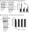
CuII(gtsm) modulates GSK3β signaling pathways and inhibits tau phosphorylation in the brains of AD mice. (A) Western blot and densitometry analyses showing CuII(gtsm) increases phosphorylation of GSK3β, Akt and ERK1/2 in brains of AD mice. ERK1/2 could not be detected in PBS extracts of mouse brains and data for ERK1/2 was therefore obtained by using cell lysis buffer as described in the methods. Data for all other proteins was obtained by using PBS extracts. (B) Western blot and densitometry analysis of brain samples shows tau phosphorylation at ser404 is decreased after treatment with CuII(gtsm) in the absence of effects on total tau, cdk5, or P35. All Western blot densitometry data are normalized for the loading control GAPDH. P values shown indicate that treatments induced significant effects compared with the sham control. Densitometry data in A and B are mean values ± SEM.
Fig. 5.
Fig. 5.
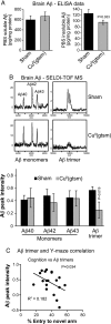
CuII(gtsm) inhibits accumulation of PBS insoluble Aβ trimers in the brains of AD mice. (A) ELISA data showing that overall levels of PBS insoluble Aβ, but not PBS soluble Aβ, are decreased in the brains of CuII(gtsm)-treated AD mice. (B) SELDI-TOF MS analysis showing Aβ trimers are decreased in the PBS-insoluble fraction of CuII(gtsm)-treated mice, but monomer levels are unaffected. All SELDI-TOF MS data are normalized for total brain protein and are expressed as Aβ peak intensity per milligram of protein. (C) Correlation analysis showing cognitive performance in Y-maze testing of AD mice improves with decreasing Aβ trimer levels in the PBS-insoluble fraction of the brain. P values shown indicate that treatments induced significant effects compared with the sham control. Data in A and Aβ peak intensity data in B are mean values ± SEM.
Fig. 6.
Fig. 6.
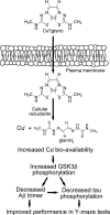
Schematic overview of proposed mechanism by which CuII(gtsm) decreases Aβ oligomer burden (trimers) and tau phosphorylation in the brains of AD mice. Cell-permeable CuII(gtsm) dissociates in the presence of intracellular reductants to release CuI and increase Cu bioavailability, thereby activating cell signaling pathways that lead to decreased Aβ trimer levels, decreased tau phosphorylation, and improved performance of AD mice in the Y-maze test for spatial learning and memory.

References

    1. Crouch PJ, et al. Mechanisms of Aβ mediated neurodegeneration in Alzheimer's disease. Int J Biochem Cell Biol. 2008;40:181–198. - PubMed
    1. Borchelt DR, et al. Familial Alzheimer's disease-linked presenilin 1 variants elevate Aβ1-42/1-40 ratio in vitro and in vivo. Neuron. 1996;17:1005–1013. - PubMed
    1. De Strooper B. Aph-1, Pen-2, and nicastrin with nresenilin generate an active γ-secretase complex. Neuron. 2003;38:9–12. - "V体育平台登录" PubMed
    1. Scheuner D, et al. Secreted amyloid β-protein similar to that in the senile plaques of Alzheimer's disease is increased in vivo by the presenilin 1 and 2 and APP mutations linked to familial Alzheimer's disease. Nat Med. 1996;2:864–870. - "VSports" PubMed
    1. Vassar R, et al. Beta-secretase cleavage of Alzheimer's amyloid precursor protein by the transmembrane aspartic protease BACE. Science. 1999;286:735–741. - PubMed

Publication types

MeSH terms

LinkOut - more resources (V体育平台登录)