V体育ios版 - Skip to main page content
U.S. flag

An official website of the United States government

Dot gov

The . gov means it’s official. Federal government websites often end in . gov or VSports app下载. mil. Before sharing sensitive information, make sure you’re on a federal government site. .

Https

The site is secure. The https:// ensures that you are connecting to the official website and that any information you provide is encrypted and transmitted securely. V体育官网.

. 2008;3(12):e4089.
doi: 10.1371/journal.pone.0004089. Epub 2008 Dec 31.

"VSports app下载" Concurrent generation of effector and central memory CD8 T cells during vaccinia virus infection

Affiliations

Concurrent generation of effector and central memory CD8 T cells during vaccinia virus infection (V体育ios版)

"VSports" Amale Laouar et al. PLoS One. 2008.

"VSports最新版本" Abstract

It is generally thought that during the contraction phase of an acute anti-viral T cell reponse, the effector T cells that escape activation-induced cell death eventually differentiate into central memory T cells over the next several weeks. Here we report that antigen-specific CD8T cells with the phenotype and function of central memory cells develop concomitantly with effector T cells during vaccinia virus (vv) infection. As soon as 5 days after an intraperitoneal infection with vv, we could identify a subset of CD44(hi) and CD62L(+) vv-specific CD8 T cells in the peritoneal exudate lymphocytes. This population constituted approximately 10% of all antigen-specific T cells and like central memory T cells, they also expressed high levels of CCR7 and IL-7R but expressed little granzyme B. Importantly, upon adoptive transfer into naïve congenic hosts, CD62L(+), but not CD62L(-) CD8 T cells were able to expand and mediate a rapid recall response to a new vv challenge initiated 6 weeks after transfer, confirming that the CD62L(+) vv-specific CD8 T cells are bonafide memory cells. Our results are thus consistent with the branched differentiation model, where effector and memory cells develop simultaneously. These results are likely to have implications in the context of vaccine design, particularly those based on vaccinia virus recombinants. VSports手机版.


Conflict of interest statement

Competing Interests: The authors have declared that no competing interests exist.

"VSports注册入口" Figures

Figure 1
Figure 1. Identification of CD44hi CD62Lhi CD8 T cell population during an acute infection with listeria monocytogenes.
C57 mice were adoptively transferred with CD8 T cells isolated from uninfected naïve P14XT-GFP mice (a, input, left panel) and infected with rLmgp33. Eight days later, PELs from infected mice were examined for the presence and phenotype of transferred cells. A representative dot plot (a, right panel) and cumulative data from 6 mice (b) of CD44 and CD62L expression by CD8 and gp33Db tetramer-gated cells is shown. The bar graph in (c) shows the mean fluorescence intensity (MFI) of GFP expression by the transferred input donor cells and CD62L+ and CD62L gp33Db tetramer+ cells in recipient mice after infection. Data are presented as mean±s.d. from two independent experiments with 3 mice per experiment.
Figure 2
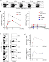
Figure 2. Kinetics and phenotype of antigen-specific CD8+ T response during an acute infection with vaccinia virus.
C57BL/6 mice were i.p. infected with vv and at different time points after infection, the presence of B8R20–27 pentamer+ CD8+ T cells in different tissues was assessed by flow cytometry. Representative dot plot of results on day 8 (a) and cumulative data from 12 mice at different time points (b) on the presence of B8R20–27 pentamer+ CD8+ T cells are shown (SPL, spleen; BM, bone marrow; PLN, peripheral (inguinal and axillaries) lymph nodes; MLN, mesenteric lymph node; Para-A-LN, Para aortic lymph node). (c) Mice were infected with vv as in (a) and at indicated times post-infection, their PELs were tested for the presence and phenotype of B8R20–27 CD8+ T cells. Representative dot plots in the left panel show the percent of B8R20–27 pentamer+ CD8+ T cells and the right panel shows CD62L and CD44 expression by the B8R20–27 pentamer+ CD8+ gated cells (n = 4). (d) shows the cumulative data on B8R20–27 pentamer+ T cells as a percent of CD8 T cells in PEL (upper panel) and CD62L+ B8R20–27 pentamer+ T-cells as a percent of total B8R+ 20–27 T-cells (lower panel).
Figure 3
Figure 3. Antigen-specific CD8+ T cells with both effector and central memory phenotype can be detected during an acute infection with vaccinia virus.
C57BL/6 mice were i.p. infected with vv and after 7 (a) or 5 (b) days of infection, the phenotype of CD62L+ and CD62L B8R20–27 CD8+ T cells in the PELs examined by flow cytometry. The indicated marker expression by CD62L+ and CD62L B8R+20–27 CD8+ gated T cells is shown. Results are representative of three independent experiments.
Figure 4
Figure 4. CD62L+ CD8 T cells isolated from vv-infected mice survive for long periods of time and mount a rapid recall response.
(a) Experimental approach for adoptive transfer experiments. PELs were harvested from day 7 or 15 vv-infected C57 (H-2Db, Thy1.2+) mice, CD8+ T cells were negatively selected and the presence of CD62L+ and CD62L B8R+ 20–27 CD8+ cells confirmed. The CD62L and CD62L+ CD8+ T-cell subsets were further isolated (purity of isolated cells is shown in the bottom dot plots) and injected into naïve sex-matched congenic recipient C57 (H-2Db, Thy1.1+) mice and the mice rested for different times before challenging with vv. (b) After 8 days of the transfer, the recipient mice were infected with vv and 3 days later, their PELs were tested for the presence of donor-derived Thy1.2+ B8R+ 20–27 CD8+ T cells (left panel) or tested for intracellular IFN-γ production after ex vivo stimulation with B8R peptide-pulsed DC (right panel). One representative result from three mice each, transferred with CD62L or CD62L+ CD8 T cells are shown. The numbers within the dot plots in the left panel represent percentage of total cells in the PEL and in the right panel, they represent percentage of cytokine+ cells among all donor cells (c) Viral titer from the ovaries of the same recipient mice in (b) is shown (n = 3). (d) Six weeks after of the transfer of cells obtained from day 15 vv-infected mice, the recipient mice were challenged with vv and after 3 days, the presence of Thy1.2+ donor-derived CD8 T cells tested in the CD62L and CD62L+ CD8 T cell transferred mice. Representative dot plot (left panel) and cumulative data from four mice on the presence of donor derived Thy1.2+ CD8 T cells are shown. The numbers within the dot plots in the left panel represent percentage of total cells in the PEL. (e) Viral titers from the ovaries of the same recipient mice in (d) is shown (n = 4). (f) After 6 weeks of the transfer of cells obtained from day 7 vv-infected mice, the recipient mice were challenged with vv and after 3 days, the presence of Thy1.2+ donor-derived CD8 T cells in the PELs were tested in the CD62L and CD62L+ CD8 T cell transferred mice. Representative dot plot (left panel) and cumulative data from four mice on the presence of donor derived Thy1.2+ CD8 T cells are shown. The numbers within the dot plots in the left panel represent percentage of total cells in the PEL. (g) IFNγ production by CD8 T cells in mice described in (f) is shown. (h) Viral titers from the ovaries of the same recipient mice in (f) is shown (n = 4).

References

    1. Butz EA, Bevan MJ. Massive expansion of antigen-specific CD8+ T cells during an acute virus infection. Immunity. 1998;8:167–175. - "V体育官网入口" PMC - PubMed
    1. Murali-Krishna K, Altman JD, Suresh M, Sourdive DJ, Zajac AJ, et al. Counting antigen-specific CD8 T cells: a reevaluation of bystander activation during viral infection. Immunity. 1998;8:177–187. - PubMed
    1. Gourley TS, Wherry EJ, Masopust D, Ahmed R. Generation and maintenance of immunological memory. Semin Immunol. 2004;16:323–333. - PubMed
    1. Antia R, Ganusov VV, Ahmed R. The role of models in understanding CD8+ T-cell memory. Nat Rev Immunol. 2005;5:101–111. - PubMed
    1. Badovinac VP, Harty JT. Programming, demarcating, and manipulating CD8+ T-cell memory. Immunol Rev. 2006;211:67–80. - PubMed

Publication types

"VSports" Substances

"V体育官网入口" LinkOut - more resources