Skip to main page content
U.S. flag

An official website of the United States government

Dot gov

The . gov means it’s official. Federal government websites often end in VSports app下载. gov or . mil. Before sharing sensitive information, make sure you’re on a federal government site. .

Https

The site is secure. The https:// ensures that you are connecting to the official website and that any information you provide is encrypted and transmitted securely V体育官网. .

. 2008 Jan;1(1):38-48.
doi: 10.1038/mi.2007.4.

Retinoic acid receptor signaling levels and antigen dose regulate gut homing receptor expression on CD8+ T cells (V体育2025版)

Affiliations

"V体育ios版" Retinoic acid receptor signaling levels and antigen dose regulate gut homing receptor expression on CD8+ T cells

M Svensson (V体育ios版) et al. Mucosal Immunol. 2008 Jan.

Abstract (VSports最新版本)

Recent studies have highlighted a central role for intestinal dendritic cells (DCs) and vitamin A metabolite retinoic acid (RA) in the generation of alpha4beta7(+) CCR9(+)"gut tropic" effector T cells VSports手机版. Here, using RA-responsive element reporter mice, we demonstrate that both splenic and mesenteric lymph node (MLN) DCs enhanced retinoic acid receptor (RAR) signaling in CD8(+) T cells; however, only a subset of MLN DCs, expressing the integrin alpha-chain CD103, induced an early RAR signal that is required for efficient CCR9 induction. MLN-primed CD8(+) T cells also received enhanced RAR-dependent signals compared with splenic-primed CD8(+) T cells in vivo. Further DC-mediated induction of gut homing receptors was inhibited at a high antigen dose without influencing RAR signaling events, and resulted in less efficient CD8(+) T-cell entry into the small intestinal mucosa. These results highlight a complex interplay between antigen dose and DC subset-induced RAR signaling events in the generation of tissue tropic effector T-cell subsets. .


Conflict of interest statement

Disclosure

The authors declare no conflict of interest.

Figures

Figure 1
Figure 1. CCR9 and α4β7 induction requires signaling via RA receptors.
(a) Experimental setup to investigate the nature of CCR9-inducing signals. (II–IV) Purified CD11c+ splenic DCs (2.5×10⁵) were pulsed with pOVA(1 nM) and incubated with 5×10⁵ CFSE-labeled OT-I cells in the lower chamber of a 0.4 µm transwell. (I, III, and IV) Purified CD11c+ MLN DCs (105) were pulsed with 1 nM pOVA and added (III) alone or (I and IV) together with 2×10⁵ CFSE-labeled OT-I cells to the upper insert. After 4 days of culture, OT-I cells cultured with (I) MLN DCs or (II–IV) splenic DCs were analyzed for CCR9 expression by flow cytometry. Results are from one representative experiment of three performed. (b and c) RAR signaling is required for induction of CCR9 and α4β7 expression on OT-I cells. Antigen-loaded (20 pM) MLN DCs (105) were co-cultured with OT-I cells (2×10⁵) in the presence or absence of pan-RAR inhibitor AGN194310. Following 4 days of culture, CCR9 and α4β7 expression was determined on the OT-I cells by flow cytometry. (c) Bars represent mean±s.e.m. from three to four experiments. *P < 0.05 compared to no added inhibitor. (d) RAR signaling is required for splenic DC-mediated induction of α4β7 expression on OT-I cells. Antigen-loaded (20 pM) splenic DCs (105) were co-cultured with OT-I cells (2×10⁵) in the presence or absence of pan-RAR inhibitor AGN194310. Following 4 days of culture, α4β7 expression was determined on the OT-I cells by flow cytometry. One representative experiment of four performed. CCR, CC chemokine receptor; CXCR, CXC chemokine receptor; DC, dendritic cell; MLN, mesenteric lymph node; pOVA, ovalbumin peptide; RA, retinoic acid; RAR, retinoic acid receptor; SPL, splenic.
Figure 2
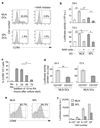
Figure 2. CD103+ MLN but not CD103 MLN or splenic DCs provide early RAR-stimulating signals required for efficient CCR9 induction.
(a) CCR9 expression by CD8+ T cells stimulated with anti-CD3 antibody in the presence of MLN or splenic DCs. A total of 2×10⁵ DR5.CD8 cells were stimulated with anti-CD3 antibody (10 µg ml−1) in the presence of 105 MLN or splenic DCs, with or without the pan-RAR inhibitor AGN194310 (100 nM). Expression of CCR9 was determined by flow cytometry after 96 h. Numbers represent percentage of CCR9+ CD8+ T cells. (b) Luciferase activity of DR5.CD8 cells stimulated with anti-CD3 antibody in the presence or absence of MLN or splenic DCs. DR5.CD8 cells were cultured as in a. Luciferase activity was measured after 24 h (upper panel) or 72 h (lower panel). Bars represent mean±s.e.m. from four to six culture wells from two to three experiments. *P < 0.05, **P < 0.01. (c) Early RAR signaling is required for efficient CCR9 induction in responding T cells. CD8+ T cells were stimulated with anti-CD3 antibody (10 µg ml−1). RA (10 nM) was added at indicated time points, and T-cell expression of CCR9 determined after 96 h of culture. Bars represent mean±s.e.m. of the percentages CCR9+ OT-I cells from four individual wells from two experiments. *P < 0.05. (d) Luciferase activity of anti-CD3 antibody (10 µg ml−1)-stimulated DR5.CD8 cells cultured in the presence of 105 CD103+ or CD103 MLN DCs isolated from Flt3L-treated mice. Luciferase activity was measured after 24 and 72 h and was normalized to the protein content of each individual well. Bars represent mean±s.e.m. from five to eight wells from two experiments. **P < 0.01. (e and f) Luciferase activity of in vivo-activated CD8+ T cells. DR5.OT-I mice were injected with OVA (200 µg, IP). Forty-two hours later, CD8+ T cells were purified from the spleen and MLN by MACS and analyzed for (e) CD69 expression by flow cytometry and (f) luciferase activity. (e) Numbers represent percentage of positive cells. (f) Bars represent mean±s.d. of three wells. CCR, CC chemokine receptor; DC, dendritic cell; IP, intraperitoneally; MACS, magnetic cell sorting; MLN, mesenteric lymph node; OVA, ovalbumin; RA, retinoic acid; RAR, retinoic acid receptor; SPL, splenic.
Figure 3
Figure 3. Induction of CCR9, α4β7, and E-selectin ligand expression in vitro is antigen dose dependent.
(ac) Purified CD11c+ MLN or splenic DCs (105) were pulsed with pOVA and incubated with 2×10⁵ CFSE-labeled OT-I cells. After 4 days of culture, OT-I cells were analyzed for expression of (ac) CCR9 and (b and c) α4β7 by flow cytometry. (b) Bars represent mean±s.e.m. of the percentage positive OT-I cells (n = 6). **P < 0.01. (c) OT-I cells were analyzed for CCR9 and α4β7 expression as a function of division. Mean±s.e.m. from six experiments. *P < 0.05, **P < 0.01; asterisks are placed between compared groups. (d) Induction of CCR9 and E-selectin ligand expression by CD103+ and CD103 MLN DCs is antigen dose dependent. Purified CD103+ and CD103 CD11c+ MLN DCs (105) were pulsed with pOVA and incubated with 2×10⁵ CFSE-labeled OT-I cells. OT-I cells were analyzed for CCR9 and E-selectin ligand expression after 4 and 6 days of culture, respectively. Bars represent mean±s.e.m. of the percentage positive OT-I cells (n = 3–7). *P < 0.05. CCR, CC chemokine receptor; DC, dendritic cell; MLN, mesenteric lymph node; pOVA, ovalbumin peptide; SPL, splenic.
Figure 4
Figure 4. High antigen dose inhibition of RAR-mediated CCR9 expression does not affect RAR signaling events.
(a) CD11c+ splenic DCs (105) were pulsed with pOVA and incubated with 2×10⁵ CFSE-labeled OT-I cells in the presence of indicated concentrations of RA. After 4 days of culture, OT-I cells were analyzed for CCR9 expression. Numbers are percentage of CCR9+ -responding OT-I cells. Results are from one representative experiment of four to five performed. (b) RAR signaling activity does not decrease with increasing antigen dose. A total of 2×10⁵ CD8+ DR5.OT-I cells were activated by 105 MLN DCs pulsed with various doses of pOVA, and luciferase activity was measured after 24 h. Data are normalized to the luciferase activity in cells stimulated by MLN DCs pulsed with 20 pM pOVA and have been normalized for variation in protein content. Bars represent mean±s.e.m. from four to six culture wells from two to three experiments. Freshly isolated unstimulated DR5.OT-I cells (5×10⁵) show < 10 luciferase (a.u.) per 5 µg protein. CCR, CC chemokine receptor; DC, dendritic cell; MLN, mesenteric lymph node; pOVA, ovalbumin peptide; RA, retinoic acid; RAR, retinoic acid receptor.
Figure 5
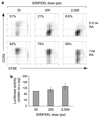
Figure 5. CCR9 induction in vivo is antigen dose dependent.
(ac) OT-I cells (3–5×10⁶) were labeled with CFSE and transferred IV into C57BL/6-CD45.1 recipient mice, and OVA was administered IP at the doses indicated. Mice were killed 2 or 3 days later, and CCR9 expression was determined by flow cytometry. (a) Numbers represent the percentage of CCR9+ cells among responding OT-I cells. (b) Bars represent the mean±s.e.m. of the percentage CCR9+ OT-I cells (n = 2–6). **P < 0.01. (c) CCR9 expression as a function of cell division. Bars represent mean±s.e.m. of the percentage CCR9+ OT-I cells after indicated number of cell divisions (n = 2–4). *P < 0.05. (d and e) OT-I cells primed by a high antigen dose have reduced capacity to localize to the small intestinal epithelium. OT-I cells were primed in vitro with low or high antigen dose-pulsed MLN DCs and after 7 days mixed at an equal ratio and injected IV into C57BL/6-CD45.1 recipient mice. The ratio of low dose-primed OT-I cells (CD45.1+ CD45.2+) and high dose-primed OT-I cells (CD45.1 CD45.2+) was assessed in the spleen, MLN, and small intestinal epithelium after 24 h by flow cytometry. (d) Representative flow cytometry analysis of transferred OT-I cells in the spleen, MLN, and small intestinal epithelium after gating on donor cells. (e) Bars represent mean±s.d. of the ratio of low-dose/high-dose-generated cells after normalization to the input ratio (n = 3). CCR, CC chemokine receptor; DC, dendritic cell; IP, intraperitoneally; IV, intravenously; MLN, mesenteric lymph node; OVA, ovalbumin; SIEP, small intestinal epithelium; SPL, spleen.

References (VSports注册入口)

    1. Briskin MD, Winsor-Hines A, Shyjan N, Cochran S, Bloom J, Wilson LM, et al. Human mucosal addressin cell adhesion molecule-1 is preferentially expressed in intestinal tract and associated lymphoid tissue. Am J Pathol. 1997;151:97–110. - PMC - PubMed
    1. Kunkel EJ, Campbell JJ, Haraldsen G, Pan J, Boisvert J, Roberts AI, et al. Lymphocyte CC chemokine receptor 9 and epithelial thymus-expressed chemokine (TECK) expression distinguish the small intestinal immune compartment: epithelial expression of tissue-specific chemokines as an organizing principle in regional immunity. J Exp Med. 2000;192:761–768. - PMC - PubMed
    1. Svensson M, Marsal J, Ericsson A, Carramolino L, Broden T, Marquez G, et al. CCL25 mediates the localization of recently activated CD8alphabeta(+) lymphocytes to the small-intestinal mucosa. J Clin Invest. 2002;110:1113–1121. - "VSports在线直播" PMC - PubMed
    1. Zabel BA, Agace WW, Campbell JJ, Heath HM, Parent D, Robert AI, et al. Human G protein-coupled receptor GPR-96/CC chemokine receptor 9 is selectively expressed on intestinal homing T lymphocytes, mucosal lymphocytes, and thymocytes and is required for thymus-expressed chemokine-mediated chemotaxis. J Exp Med. 1999;190:1241–1256. - PMC - PubMed
    1. Wurbel MA, Philippe JM, Nguyen C, Victorero G, Freeman T, Wooding P, et al. The chemokine TECK is expressed by thymic and intestinal epithelial cells and attracts double- and single-positive thymocytes expressing the TECK receptor CCR9. Eur J Immunol. 2000;30:262–271. - PubMed

V体育官网 - Publication types

MeSH terms