Skip to main page content
U.S. flag

An official website of the United States government

Dot gov

The . gov means it’s official. Federal government websites often end in . gov or . mil. Before sharing sensitive information, make sure you’re on a federal government site. VSports app下载.

Https

The site is secure. The https:// ensures that you are connecting to the official website and that any information you provide is encrypted and transmitted securely V体育官网. .

. 2009 Sep;29(8):1253-61.
doi: 10.1111/j.1478-3231.2008.01921.x. Epub 2008 Nov 15.

Cholangiocytes as immune modulators in rotavirus-induced murine biliary atresia

Affiliations

"VSports最新版本" Cholangiocytes as immune modulators in rotavirus-induced murine biliary atresia

Barrett H Barnes et al. Liver Int. 2009 Sep.

"VSports在线直播" Abstract

Background/aims: Biliary atresia (BA) is a progressive disease characterized by bile duct inflammation and fibrosis. The aetiology is unknown and may be due to a virus-induced, autoimmune-mediated injury of cholangiocytes VSports手机版. Cholangiocytes are not only targets of injury but may also modulate hepatic inflammation. The aim of this study was to determine the immune profile of murine cholangiocytes and the ability to function as antigen-presenting cells (APCs) in culture with Rhesus rotavirus (RRV), poly I:C (viral mimic) or interferon-gamma/tumour necrosis factor-alpha. .

Methods/results: Both the cholangiocyte cell line (long-term culture) and fresh, ex vivo cholangiocytes expressed APC surface markers major histocompatibility complex (MHC)-class I and II and CD40, while only the cultured cell line expressed costimulatory molecules B7-1 and B7-2. Despite APC expression, cultured cholangiocytes were unable to function as competent APCs in T-cell proliferation assays. Furthermore, both cultured and ex vivo cholangiocytes expressed RNA transcripts for many pro-inflammatory cytokines and chemokines. V体育安卓版.

Conclusions: Although cholangiocytes contain APC molecules, they are incompetent at antigen presentation and cannot elicit effective T-cell activation. Upregulation of MHC-class I and II found in BA mice may serve to prime the cholangiocyte as a target for immune-mediated injury V体育ios版. Cholangiocytes produced many pro-inflammatory cytokines and chemokines in the setting of RRV infection and T-helper type 1 cytokine milieu, suggesting a role of cholangiocytes as immune modulators promoting the ongoing inflammation that exists in RRV-induced BA. .


VSports app下载 - Figures
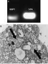
Fig. 1
Fig. 1
Rhesus rotavirus (RRV) infection of cholangiocytes. (A) Cultured cholangiocytes infected with RRV (multiplicity of infection of 50, 3 h) contained mRNA transcripts for nonstructural protein 1 (NSP1) and viral structural protein 4 (VP4). (B) Electron microscopy of RRV-infected cholangiocytes revealed intracytoplasmic RRV particles (black arrows).
Fig. 2
Fig. 2
Fluorescent-activated cell sorting (FACS) analysis of antigen-presenting cell (APC) surface markers on the cultured cholangiocyte cell line. (A) The cultured cholangiocyte cell line was incubated with fluorescein isothiocyanate-labelled antibodies to major histocompatibility complex (MHC) I, MHC II, CD40, CD80 or CD86 followed by FACS analysis that revealed cell surface expression of all APC markers (solid grey) compared with isotype controls (thin black line). (B) Cultured cholangiocytes infected with RRV (multiplicity of infection of 50, 24 h) expressed higher levels of CD86 (heavy black line) compared with uninfected controls (solid grey) and isotype controls (thin black line). (C) Cultured cholangiocytes treated with interferon-γ and tumour necrosis factor-α showed marked increase in MHC I expression (heavy black line) compared with uninfected controls (solid grey) and isotype controls (thin black line).
Fig. 3
Fig. 3
Antigen-presenting cell (APC) surface markers on cholangiocytes isolated ex vivo from livers of balanced salt solution (BSS) control and Rhesus rotavirus (RRV)-induced biliary atresia (BA) mice. (A) Cholangiocytes were identified based on cell surface expression of TROMA III (anticytokeratin antibody). The specificity of this antibody was confirmed by immunohistochemistry staining of mouse liver and bile duct tissue. On the left is an extrahepatic bile duct incubated with anti-TROMA III-AF555 (red) and counterstained with 4,6-diamidino-2-phenylindole nuclear stain (blue) (magnification: × 200). On the right, fluorescent-activated cell sorting analysis of liver cells revealed that approximately 5–7% of all liver cells were cholangiocytes (TROMA III positive). (B) Ex vivo cholangiocytes from BSS and BA mice were double stained with TROMA III-AF555 and various APC markers-fluorescein isothiocyanate (FITC) and analysed by flow cytometry. Normal cholangiocytes express major histocompatibility complex (MHC) class I and CD40 while cholangiocytes from BA mice showed upregulation of these APC molecules as well as aberrant expression of MHC class II in approximately 16% of the population. Isotype controls were negative (data not shown).
Fig. 4
Fig. 4
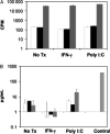
Functional antigen presentation of cultured cholangiocytes as measured by lymphoproliferation assay and interleukin (IL)-2 production. (A) Cholangiocytes functioned as experimental antigen-presenting cells (APCs) and were pretreated with media alone, poly I:C or interferon-γ (IFN-γ) before mitomycin C treatment. 2.5 × 10⁵ cholangiocytes were cultured with 2.5 × 10⁵ DO11.10 T cells in the absence (white bars) or presence (black bars) of 1 μg/ml ovalbumin (OVA)323–339 antigen. As a positive control, DO11.10 T cells were cultured with 2.5 × 10⁵ mitomycin C-treated bulk splenocytes with OVA antigen (grey bar). Cultures were pulsed with 1 μCi/well of thymidine-[methyl-3H] at 72 h and harvested 18 h thereafter. Values represent c.p.m. ± SEM of triplicate cultures. Pretreated cholangiocytes were unable to induce OVA antigen-specific T-cell proliferation compared with control APCs. (B) Cholangiocytes were pretreated with media alone, poly I:C or IFN-γ before mitomycin C treatment. 2.5 × 10⁵ cholangiocytes were cultured alone (white bar), or cocultured with 2.5 × 10⁵ OVA-specific DO11.10 hybridoma cells and without (black bars) or with OVA antigen (grey bars). As a positive control, DO11.10 hybridoma cells were cultured with 2.5 × 10⁵ mitomycin C-treated bulk splenocytes with OVA antigen (hatched bar). Culture supernatants were collected at 24 h and analysed by enzyme-linked immunosorbent assay for IL-2 protein. Values represent the mean pg/ml ± SEM. Cholangiocytes were unable to induce IL-2 secretion from DO11.10 hybridomas in the presence of OVA antigen compared with control APCs.
Fig. 5
Fig. 5
Cultured cholangiocytes produce pro-inflammatory cytokines and chemokines. RNA was extracted from the cholangiocyte cell line alone (A) or cholangiocytes pretreated with Rhesus rotavirus (RRV) (B), poly I:C (C) or interferon-γ (IFN-γ) (D). RT-PCR was performed on cDNA using interleukin (IL)-1β, IL-6, tumour necrosis factor-α (TNF-α), IFN-γ, transforming growth factor-β (TGF-β), monokine induced by γ-interferon (MIG), macrophage inflammatory protein-1α (MIP-1α), γ-interferon inducible protein-10 (IP-10) and regulated upon activation, normal T-cell expressed and secreted (RANTES) primer pairs.
Fig. 6
Fig. 6
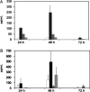
Cultured cholangiocytes produce interleukin (IL)-6 and transforming growth factor-β (TGF-β) proteins. Cholangiocytes from the cell line were cultured alone (white bar) or with interferon-γ (IFN-γ) (black bar), poly I:C (grey bar) or Rhesus rotavirus (hatched bar) and culture supernatants were collected at 24, 48 and 72 h to assess for IL-6 (A) and TGF-β (B) proteins by enzyme-linked immunosorbent assay. Values represent the mean pg/ml ± SEM. Cholangiocytes treated with poly I:C generated modest amounts of IL-6 and those treated with IFN-γ produced abundant amounts of IL-6 and TGF-β compared with unstimulated controls at 24 and 48 h in culture.
Fig. 7
Fig. 7
Cytokine and chemokine profile of purified cholangiocytes isolated ex vivo from murine balanced salt solution (BSS) control and Rhesus rotavirus (RRV)-induced biliary atresia (BA) mice. Cholangiocytes were isolated by magnetic cell sorting (purity > 98%) from pooled liver samples of either BSS control or BA mice. RNA was extracted and RT-PCR was performed on cDNA using interleukin (IL)-1β, IL-6, tumour necrosis factor-α (TNF-α), interferon-γ (IFN-γ), transforming growth factor-β (TGF-β), monokine induced by γ-interferon (MIG), macrophage inflammatory protein-1α (MIP-1α), γ-interferon inducible protein-10 (IP-10) and regulated upon activation, normal T-cell expressed and secreted (RANTES) primer pairs.

References

    1. Chardot C, Carton M, Spire-Bendelac N, et al. Epidemiology of biliary atresia in France: a national study 1986–96. J Hepatol. 1999;31:1006–13. - "VSports" PubMed
    1. Chen SM, Chang MH, the Taiwan Infant Stool Color Card Study Group et al. Screening for biliary atresia by infant stool color card in Taiwan. Pediatrics. 2006;117:1147–54. - PubMed
    1. Karrer FM, Price MR, Bensard DD, et al. Long-term results with the Kasai operation for biliary atresia. Arch Surg. 1996;131:493–6. - PubMed
    1. Karrer FM, Bensard DD. Neonatal cholestasis. Semin Pediatr Surg. 2000;9:166–9. - PubMed
    1. Mack CL, Sokol RJ. Unraveling the pathogenesis and etiology of biliary atresia. Pediatr Res. 2005;57:87R–94R. - PubMed

Publication types

MeSH terms

Substances