V体育2025版 - Skip to main page content
U.S. flag

An official website of the United States government

Dot gov

The . gov means it’s official VSports app下载. Federal government websites often end in . gov or . mil. Before sharing sensitive information, make sure you’re on a federal government site. .

Https

The site is secure V体育官网. The https:// ensures that you are connecting to the official website and that any information you provide is encrypted and transmitted securely. .

. 2008 Dec;10(12):1477-83.
doi: 10.1038/ncb1807. Epub 2008 Nov 23.

Glucose metabolism inhibits apoptosis in neurons and cancer cells by redox inactivation of cytochrome c

Affiliations

V体育2025版 - Glucose metabolism inhibits apoptosis in neurons and cancer cells by redox inactivation of cytochrome c

Allyson E Vaughn et al. Nat Cell Biol. 2008 Dec.

Abstract

Neurons and cancer cells use glucose extensively, yet the precise advantage of this adaptation remains unclear. These two seemingly disparate cell types also show an increased regulation of the apoptotic pathway, which allows for their long-term survival. Here we show that both neurons and cancer cells strictly inhibit cytochrome c-mediated apoptosis by a mechanism dependent on glucose metabolism. We report that the pro-apoptotic activity of cytochrome c is influenced by its redox state and that increases in reactive oxygen species (ROS) following an apoptotic insult lead to the oxidation and activation of cytochrome c VSports手机版. In healthy neurons and cancer cells, however, cytochrome c is reduced and held inactive by intracellular glutathione (GSH), generated as a result of glucose metabolism by the pentose phosphate pathway. These results uncover a striking similarity in apoptosis regulation between neurons and cancer cells and provide insight into an adaptive advantage offered by the Warburg effect for cancer cell evasion of apoptosis and for long-term neuronal survival. .


Figures

Figure 1
Figure 1. Endogenous cytochrome c release is incapable of inducing apoptosis in NGF-maintained sympathetic neurons
a) Cultures of MEFs or sympathetic neurons were injected with tBid-GFP, or GFP plasmid. Cell survival was quantified by cell morphology and expressed as a percentage of alive and healthy green cells at 24 hrs compared to 5 hrs post-injection. *represents no MEF survival. Representative photographs of MEFs and neurons are shown; arrows point to injected cells. b) XIAP -/- sympathetic neurons or wildtype littermates were injected with EGFP or tBid-GFP, and cell survival was quantified as in (a). c) tBid-GFP was injected into sympathetic neurons, and allowed to express for 24 hrs. Cells were fixed, and the status of cytochrome c analyzed by immunofluorescence. Percentage of neurons with mitochondrial cytochrome c is quantified in XIAP -/- neurons. d) XIAP -/- sympathetic neurons were either maintained in NGF-containing media, or deprived of NGF for 8 hrs (with or without 50 μM zVAD), followed by injection with EGFP or tBid-GFP. Cell survival was quantified by cell morphology and expressed as a percentage of alive and healthy green cells at 16 hrs compared to 5 hrs post-injection. e) XIAP -/- sympathetic neurons were deprived of NGF (with or without 50 μM zVAD) for 12 hrs followed by injection with rhodamine alone, or cytochrome c (2.5 μg/μl) along with rhodamine to mark injected cells. Cell survival was assessed at various time points following injection. Error bars represent ±SEM of three independent experiments.
Figure 2
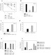
Figure 2. Oxidation of cytochrome c increases its apoptotic activity
a) XIAP -/- sympathetic neurons were treated with 20-50 μM of H2O2 for 20 minutes, followed by injection with cytochrome c protein and rhodamine, or rhodamine dye alone. Cell survival was assessed at various time points following injection. b) XIAP -/- sympathetic neurons were treated with 10 mM GSH ethyl ester for 12 hrs, followed by injection of cytochrome c along with rhodamine, or rhodamine alone. Cell survival was assessed at 16 hrs following injection. c) XIAP -/- sympathetic neurons were injected with either cytochrome c, or cytochrome c that was pre-incubated with 10 Units/ml of cytochrome c reductase. Cell survival was assessed at 16 hrs following injection. d) XIAP -/- sympathetic neurons were injected with either cytochrome c, or cytochrome c that had been pre-incubated with DTT (and subsequently separated) to reduce this cytochrome c. Percent survival was assessed after 6 hrs. e) XIAP -/- sympathetic neurons were deprived on NGF for 8 hrs in the presence of 10 mM GSH, followed by injection with EGFP or tBid-GFP constructs. Cell survival was quantified by cell morphology and expressed as a percentage of alive and healthy green cells at 16 hrs compared to 5 hrs post-injection. f) Exogenous cytochrome c (which is primarily oxidized) was added at a concentration of 10 uM to neuronal extracts, and Abs550 was measured following a 15 min. incubation. Control experiments measuring Abs550 of reduced or oxidized cytochrome c are also shown. Results are mean (±SEM) of three independent experiments.
Figure 3
Figure 3. Role of the pentose phosphate shunt in cytochrome c-mediated apoptosis
a) Average ROS levels in sympathetic neurons were observed by fluorescence intensity of the redox-sensitive dye CM-H2DCFDA following 30 min of GSH depletion with 0.1 mM DEM. b) XIAP -/- sympathetic neurons (NGF-maintained) were treated with 0.1 mM DEM for 30 min followed by injection of cytochrome c and rhodamine, or rhodamine dye alone. Cell survival was assessed at various timepoints. c) Average ROS levels were observed as in (a) following inhibition of the Pentose Phosphate Pathway with 200 μM DHEA for 24 hrs. d) XIAP -/- sympathetic neurons (NGF-maintained) were treated with 200 μM DHEA for 6 hrs, 0.1 mM 6-AN for 24 hrs, or left untreated, followed by injection with tBid-GFP or EGFP constructs. Cell survival was expressed as a percentage of healthy green cells at 16 hrs compared to 5 hrs post-injection. Error bars represent ±SEM of n≥3.
Figure 4
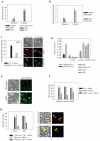
Figure 4. Glucose metabolism protects cancer cells from cytochrome c-mediated apoptosis
a) Normal cells (Human Mammary Epithelial Cells-HuMECs, MEFs, Human Dermal Fibroblasts-HDFs) or cancer cell lines (Sk-Mel 103, HeLa, JM2) were injected with cytochrome c and Smac protein and cell survival was assessed after 30 minutes. b) Total glutathione (GSH) was measured in normal mitotic cells as well as cancer cell lines, and expressed as a concentration of GSH to total cellular protein. c) MEFs were treated with 5 mM GSH ethyl ester for 15 min, followed by injection with cytochrome c. After 1 hr, injected cells were assessed for Annexin V positivity. Photographs show representative Annexin V-FITC staining of cytochrome c injected cells (arrows). d) Exogenous cytochrome c (which is primarily oxidized) was added at a concentration of 10 μM to normal or cancer cell extract, and Abs550 was measured following a 15 min incubation. e) Average ROS levels in HeLa cells measured by fluorescence of CMH2DCFDA in the absence or presence of the pentose phosphate pathway inhibitor, DHEA (200 μM) for 6 hrs. f) The pentose phosphate pathway was inhibited in various cancer cell lines for 6 hrs by addition of 200 μM DHEA, followed by injection of cytochrome c and Smac protein. Cell survival was assessed at 3 hrs. g) JM2 and HeLa cells were deprived of glucose for 16 hrs followed by injection with cytochrome c and Smac, and assessed for survival at various time points. Images are representative of JM2 cells at 3 hrs following cytochrome c injection. Error bars represent ±SEM of n≥3.

References (VSports app下载)

    1. Wright KM, Deshmukh M. Restricting apoptosis for postmitotic cell survival and its relevance to cancer. Cell Cycle. 2006;5:1616–1620. - PubMed
    1. Yuan J, Yankner BA. Apoptosis in the nervous system. Nature. 2000;407:802–809. - PubMed
    1. Green DR, Evan GI. A matter of life and death. Cancer Cell. 2002;1:19–30. - PubMed
    1. Wang X. The expanding role of mitochondria in apoptosis. Genes Dev. 2001;15:2922–2933. - PubMed
    1. Schubert D. Glucose metabolism and Alzheimer's disease. Ageing Res Rev. 2005;4:240–257. - PubMed

Publication types

MeSH terms