<u lang="srstoqK"></u><center lang="6bjcei"></center>
Skip to main page content
U.S. flag

An official website of the United States government

Dot gov

The VSports app下载. gov means it’s official. Federal government websites often end in . gov or . mil. Before sharing sensitive information, make sure you’re on a federal government site. .

Https

The site is secure. The https:// ensures that you are connecting to the official website and that any information you provide is encrypted and transmitted securely. V体育官网.

. 2008 Nov 19;28(47):12570-80.
doi: 10.1523/JNEUROSCI.4048-08.2008.

Neuronal death resulting from targeted disruption of the Snf2 protein ATRX is mediated by p53

Affiliations

Neuronal death resulting from targeted disruption of the Snf2 protein ATRX is mediated by p53

Claudia Seah et al. J Neurosci. .

Abstract

ATRX, a chromatin remodeling protein of the Snf2 family, participates in diverse cellular functions including regulation of gene expression and chromosome alignment during mitosis and meiosis. Mutations in the human gene cause alpha thalassemia mental retardation, X-linked (ATR-X) syndrome, a rare disorder characterized by severe cognitive deficits, microcephaly and epileptic seizures VSports手机版. Conditional inactivation of the Atrx gene in the mouse forebrain leads to neonatal lethality and defective neurogenesis manifested by increased cell death and reduced cellularity in the developing neocortex and hippocampus. Here, we show that Atrx-null forebrains do not generate dentate granule cells due to a reduction in precursor cell number and abnormal migration of differentiating granule cells. In addition, fewer GABA-producing interneurons are generated that migrate from the ventral telencephalon to the cortex and hippocampus. Staining for cleaved caspase 3 demonstrated increased apoptosis in both the hippocampal hem and basal telencephalon concurrent with p53 pathway activation. Elimination of the tumor suppressor protein p53 in double knock-out mice rescued cell death in the embryonic telencephalon but only partially ameliorated the Atrx-null phenotypes at birth. Together, these findings show that ATRX deficiency leads to p53-dependent neuronal apoptosis which is responsible for some but not all of the phenotypic consequences of ATRX deficiency in the forebrain. .


"V体育官网入口" Figures

Figure 1.
Figure 1.
Expression profile of ATRX in the developing hippocampus. A, Immunofluorescence detection of ATRX in the telencephalon at E13.5 reveals that ATRX is expressed in the mitotic and ventricular layers that line the lateral ventricle and is also highly expressed in the differentiated neurons that form the cortical plate (white arrows). Scale bar, 200 μm. Expression is detected in the ventricular zone of the hippocampal primordium (B), the cortex (C) and the basal ganglia (D). Scale bar, 100 μm. E, ATRX immunofluorescence at E13.5 shows that the protein colocalizes with condensed chromosomes in the mitotic layer (E′) and to DAPI-rich heterochromatin bundles in cycling cells of the ventricular zone (E″). Scale bar, 50 μm. F, ATRX is expressed in most cells of the hippocampus at E15.5. Scale bar, 200 μm. G, In the newborn brain, ATRX expression is maintained in the hippocampus in all cell types. Scale bar, 200 μm. H, Hippocampal primordium; C, cortex; BG, basal ganglia; sub, subiculum; fi, fibrium.
Figure 2.
Figure 2.
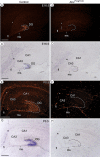
The granule cell marker Prox1 is not expressed in the ATRX-null hippocampus. Prox1 protein and transcript levels were analyzed by immunofluorescence (A, B, E, F) and by in situ hybridization (C, D, G, H), respectively. Prox1 expression was observed as early as E15.5 in the control hippocampus and can be detected in a subset of migrating precursors in the migratory stream and in differentiated granule cells that form the dentate gyrus at E16.5 and P0.5. Prox1 transcripts and protein were not detected in the ATRX-null hippocampus (AtrxFoxg1Cre). ms, Migratory stream; fi, fibrium. Asterisks indicate the site of the dentate notch. Hatched lines outline the shape of the dentate gyrus in control sections and its expected location in the AtrxFoxg1Cre sections. Scale bars, 200 μm.
Figure 3.
Figure 3.
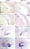
Delayed differentiation and incomplete migration of ATRX-null dentate precursors. In situ hybridization of control and AtrxFoxg1Cre sections using a DIG-labeled antisense probe specific for NeuroD1, a marker of dentate granule cell differentiation. NeuroD1 expression was detectable in the caudal and rostral hippocampal hem in control, but not AtrxFoxg1Cre sections at E13.5 (A–D, arrows). Precursor cells in both the control and ATRX-null hippocampus express NeuroD1 at E16.5 (E, F) and at P0.5 (G, H) but have not migrated to the site of the DG in the ATRX-deficient hippocampus. fi, Fibrium; ms, migratory stream. Asterisks indicate the site of the dentate notch. Hatched lines outline the shape of the dentate gyrus in control sections and its expected location in the AtrxFoxg1Cre sections. Scale bars, 200 μm.
Figure 4.
Figure 4.
Fewer precursor cells are present in the dentate neuroepithelium and the migratory stream at E16.5. Expression of Ngn2, Lef1 and Math3 that mark granule precursors was analyzed in control and AtrxFoxg1Cre hippocampus at E16.5 by in situ hybridization using specific DIG-labeled antisense probes. Fewer precursor cells are localized at the dentate notch (indicated by asterisks) and fewer Ngn2+ and Lef1+ precursors are present in the migratory stream at E16.5 (indicated by arrows). Scale bar, 200 μm.
Figure 5.
Figure 5.
Reduced expression of GABAergic markers in the ATRX-null forebrain. A, Real-time reverse transcriptase PCR of GABAergic markers in forebrain tissue isolated from E13.5 and P0.5 control and AtrxFoxg1Cre littermate embryos (n = 3). B, Immunofluorescence detection of Npy in the ventral telencephalon at E13.5 and E16.5 shows reduced staining levels in the ATRX-null embryos. Npy, Neuropeptide Y; Sst, somatostatin; Dlx5, distal-less homeobox 5; Gbx2, gastrulation brain homeobox 2; Lhx6, LIM homeobox protein 6; Cck, cholecystokinin. E13.5: Scale bar, 200 μm; E16.5: Scale bar, 150 μm.
Figure 6.
Figure 6.
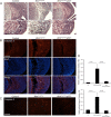
Rescue of neuronal apoptosis in the ATRX-null telencephalon by loss of p53. A, H&E staining of the hippocampal hem at E13.5 reveals pyknotic clusters in the AtrxFoxg1Cre embryos but not in control and AtrxFoxg1Cre:p53−/− embryos. Bottom micrographs show higher magnification of the regions boxed in the top micrographs. Scale bar, 50 μm. B, Immunofluorescence detection of activated caspase 3, a marker of apoptotic cell death, in the hippocampal hem at E13.5. DAPI counterstaining of the sections outlines the nuclei. Apoptotic cells are more frequently observed in AtrxFoxg1Cre compared with control and AtrxFoxg1Cre:p53−/− sections. Scale bar, 100 μm. C, Activated caspase 3 immunofluorescence detection in the basal telencephalon at E13.5 shows increased levels of apoptosis in the AtrxFoxg1Cre compared with control and AtrxFoxg1Cre:p53−/− embryos. Scale bar, 100 μm. D, Quantification of activated caspase 3 staining in the hippocampal hem and in the basal ganglia confirms a statistically significant difference in cell death in the AtrxFoxg1Cre:p53−/− embryos compared with AtrxFoxg1Cre embryos.
Figure 7.
Figure 7.
Minimal size recovery of the E16.5 Atrx-null forebrain in the absence of p53. A, H&E sections of E16.5 forebrain shows that loss of p53 does not ameliorate forebrain size, except in the caudal-medial area (indicated by *). Scale bar, 500 μm. B, Measurements of cortical plate thickness, brain height and hippocampal area confirm size reductions in the AtrxFoxg1Cre embryos but show no significant amelioration in the AtrxFoxg1Cre:p53−/− embryos. C, Partial recovery of medial cortical size is suggested by the decreased distance between the left and right cingulate cortex in the AtrxFoxg1Cre:p53−/− compound mutants compared with the AtrxFoxg1Cre embryos (* in A). H, Hippocampus; Cx, cortex. n = 3 for all genotypes except for AtrxFoxg1Cre:p53+/+ where n = 2. All distances are measured in micrometers unless otherwise indicated.
Figure 8.
Figure 8.
Partial rescue of ATRX-null forebrain size upon loss of p53 at E18.5 and P0.5. A, H&E sections of E18.5 forebrain showing partial size recovery of the hippocampus and cortex in the AtrxFoxg1Cre:p53+/− and AtrxFoxg1Cre:p53−/− compared with AtrxFoxg1Cre embryos. B, Size differences were measured as indicated in the inset (parameters 1–3) in two different caudal areas of the forebrain (C) Partial forebrain size recovery was also observed at P0.5 (top). Higher magnification micrographs show minimal rescue of cellularity in the dentate gyrus in the AtrxFoxg1Cre:p53−/− compound mutants (middle), and accumulation of dentate precursors in the dentate neuroepithelium (bottom, arrows). Scale bar, 100 μm. H, Hippocampus; Cx, cortex.

References

    1. Abidi F, Schwartz CE, Carpenter NJ, Villard L, Fontés M, Curtis M. Carpenter-Waziri syndrome results from a mutation in XNP. Am J Med Genet. 1999;85:249–251. - PubMed
    1. Altman J, Bayer SA. Migration and distribution of two populations of hippocampal granule cell precursors during the perinatal and postnatal periods. J Comp Neurol. 1990a;301:365–381. - PubMed
    1. Altman J, Bayer SA. Mosaic organization of the hippocampal neuroepithelium and the multiple germinal sources of dentate granule cells. J Comp Neurol. 1990b;301:325–342. - PubMed
    1. Bagri A, Gurney T, He X, Zou YR, Littman DR, Tessier-Lavigne M, Pleasure SJ. The chemokine SDF1 regulates migration of dentate granule cells. Development. 2002;129:4249–4260. - "V体育平台登录" PubMed
    1. Bérubé NG, Smeenk CA, Picketts DJ. Cell cycle-dependant phosphorylation of the ATRX protein correlates with changes in nuclear matrix and chromatin association. Human Molecular Genetics. 2000;9:539–547. - PubMed

"VSports在线直播" Publication types

MeSH terms