<code lang="AJnOBd"></code> Skip to main page content
U.S. flag

An official website of the United States government

Dot gov

The VSports app下载. gov means it’s official. Federal government websites often end in . gov or . mil. Before sharing sensitive information, make sure you’re on a federal government site. .

Https

The site is secure. The https:// ensures that you are connecting to the official website and that any information you provide is encrypted and transmitted securely V体育官网. .

. 2009 Mar 5;113(10):2191-201.
doi: 10.1182/blood-2008-07-169417. Epub 2008 Nov 14.

SCL and associated proteins distinguish active from repressive GATA transcription factor complexes

Affiliations

SCL and associated proteins distinguish active from repressive GATA transcription factor complexes

Tamara Tripic et al. Blood. .

Abstract

GATA-1 controls hematopoietic development by activating and repressing gene transcription, yet the in vivo mechanisms that specify these opposite activities are unknown. By examining the composition of GATA-1-associated protein complexes in a conditional erythroid rescue system as well as through the use of tiling arrays we detected the SCL/TAL1, LMO2, Ldb1, E2A complex at all positively acting GATA-1-bound elements examined VSports手机版. Similarly, the SCL complex is present at all activating GATA elements in megakaryocytes and mast cells. In striking contrast, at sites where GATA-1 functions as a repressor, the SCL complex is depleted. A DNA-binding defective form of SCL maintains association with a subset of active GATA elements indicating that GATA-1 is a key determinant for SCL recruitment. Knockdown of LMO2 selectively impairs activation but not repression by GATA-1. ETO-2, an SCL-associated protein with the potential for transcription repression, is also absent from GATA-1-repressed genes but, unlike SCL, fails to accumulate at GATA-1-activated genes. Together, these studies identify the SCL complex as a critical and consistent determinant of positive GATA-1 activity in multiple GATA-1-regulated hematopoietic cell lineages. .


Figures
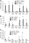
Figure 1
Figure 1
GATA-1 recruits SCL to active erythroid genes. ChIP analysis using GATA-1 (A), SCL (B), and GATA-2 (C) antibodies or isotype-matched control antibodies (IgG) and primers for indicated sites. Primers for 1 kb upstream of the Hbb-b1 promoter (−1 kb) served as negative control. ChIP experiments were performed in G1E cells (−GATA-1) and G1E-ER4 cells after E2 treatment for 21 to 24 hours (+GATA-1). The data are the averages of 3 or more independent experiments. Error bars represent SDs. *Statistical significance (P ≤ .05) based on a 2-tailed t test. Numbers indicate P values.
Figure 2
Figure 2
GATA-1, SCL, and Ldb-1 co-occupy active genes in megakaryocytes and mast cells. ChIP analysis using GATA-1, SCL, Ldb-1, or isotype-matched control antibodies in megakaryocytic L8057 cells (A) and primary bone marrow–derived mast cells (B). The −3-kb region upstream of αIIb served as negative control. The data shown are the averages of 3 or more independent experiments in panel A and 2 independent experiments in panel B. Error bars represent SDs.
Figure 3
Figure 3
Depletion of SCL from GATA-1–repressed genes. ChIP analysis using GATA-1, SCL, GATA-2, or isotype-matched control antibodies at Kit (A), Gata2 (B), the FOG-1–independent Lyl1 gene (C), and the GATA-E-box–containing genes Etv6, Sox6, and Tram2 (D). ChIP experiments were performed in parental G1E cells and estradiol-treated G1E-ER4 cells as in Figure 1. Error bars represent SDs. *Statistical significance (P ≤ .05) based on a 2-tailed t test. Numbers indicate P values. Please note that the difference in absolute signal intensity for the SCL ChIP between panel C and the other panels is due to the use of a different batch of SCL antibodies.
Figure 4
Figure 4
ETO-2 occupancy at GATA-1–regulated genes. ChIP analysis using ETO-2 or isotype-matched control antibodies at indicated GATA-1–activated (A) and GATA-1–repressed Kit (B) and Gata2 (C) genes. Prom indicates promoter. ChIP experiments were performed as in Figure 1. The data are the averages of 3 or more independent experiments. Error bars represent SDs.
Figure 5
Figure 5
SCL recruitment in the absence of DNA binding. Anti-HA or control IgG ChIP of GATA-1–activated (A) and GATA-1–repressed (B) genes in G1E-ER4 cells expressing HA-tagged SCL (HA-SCL) or HA-SCL-RER (HA-SCL*), before and after estradiol (E2) treatment for 21 to 24 hours. The data shown are the averages of 2 (Klf1, Gata2 −2.8), 3 (HS3, Hba-a1, −31, Hba-a1 promoter, Lyl1 and Rgs18 promoters), and 4 (HS2, Hbb-b1, Hbb-b1 promoter, Band3, Band3 promoter, Kit −114, Kit +5) independent experiments. Error bars represent SDs. Note that the decrease in HA-SCL occupancy at the Lyl1 gene is less pronounced than that observed with endogenous SCL (Figure 3C). This is likely the result of leakiness with regard to GATA-1-ER activity in this experiment.
Figure 6
Figure 6
Erythroid enhancers are co-occupied by GATA-1, SCL, and Ldb1. Sixty-one DNA segments occupied in vivo by GATA-1, whose enhancer activities were determined by Cheng et al, were evaluated for co-occupancy by SCL and Ldb1, based on the ChIP-on-chip results. The distribution of results after transient transfection of K562 cells (range of 8 to 24 determinations for each GATA-1–occupied DNA segment) is shown as a box plot, with the internal line indicating the median, the box extending to the first and third quartiles, and the whiskers extending to the most extreme data point that is no more than 1.5 times the interquartile range. Black boxes represent DNA segments co-occupied by GATA-1, SCL, and Ldb1; white boxes represent sequences that are occupied only by GATA-1. The gray box represents a segment that was not called as an SCL peak in ChIP-on-chip but was shown by quantitative PCR (qPCR) to be occupied by SCL. The horizontal line demarcates the threshold for enhancer activity. The results were partitioned into DNA segments with no enhancer activity (class I), activity clustering around the threshold (class II), and more than 2-fold activity (class III; see bottom panel for a summary of the results). Class I and class III are statistically different, tested by both χ2 test and Fisher exact test (P < .001).
Figure 7
Figure 7
LMO2 is required for activation but not repression of GATA-1–regulated genes. (A) Anti-LMO2 Western blot of nuclear extracts from Bcl-XL–expressing G1E-ER4 cells transduced with vector producing an shRNA against LMO2 or empty vector (ctr). β-Actin served as loading control. (B,C) Cells were treated with estradiol (E2) for 24 hours and mRNA levels of indicated genes were determined by real-time RT-PCR. Results were normalized to Gapdh mRNA and plotted as fold change compared with untreated cells. The data are the averages of 4 independent experiments. Error bars represent SD.

VSports - References

    1. Fujiwara Y, Browne CP, Cunniff K, Goff SC, Orkin SH. Arrested development of embryonic red cell precursors in mouse embryos lacking transcription factor GATA-1. Proc Natl Acad Sci U S A. 1996;93:12355–12358. - PMC - PubMed
    1. Shivdasani RA, Fujiwara Y, McDevitt MA, Orkin Stuart H. A lineage-selective knockout establishes the critical role of transcription factor GATA-1 in megakaryocyte growth and platelet development. EMBO J. 1997;16:3965–3973. - PMC - PubMed
    1. Yu C, Cantor AB, Yang H, et al. Targeted deletion of a high-affinity GATA-binding site in the GATA-1 promoter leads to selective loss of the eosinophil lineage in vivo. J Exp Med. 2002;195:1387–1395. - PMC - PubMed
    1. Migliaccio AR, Rana RA, Sanchez M, et al. GATA-1 as a regulator of mast cell differentiation revealed by the phenotype of the GATA-1low mouse mutant. J Exp Med. 2003;197:281–296. - PMC - PubMed
    1. Nishiyama C, Ito T, Nishiyama M, et al. GATA-1 is required for expression of FcεRI on mast cells: analysis of mast cells derived from GATA-1 knockdown mouse bone marrow. Int Immunol. 2005;17:847–856. - PubMed (V体育官网入口)

Publication types

MeSH terms

Substances