Skip to main page content
U.S. flag

An official website of the United States government

Dot gov

The VSports app下载. gov means it’s official. Federal government websites often end in . gov or . mil. Before sharing sensitive information, make sure you’re on a federal government site. .

Https

The site is secure. The https:// ensures that you are connecting to the official website and that any information you provide is encrypted and transmitted securely. V体育官网.

. 2008 Nov;48(5):1632-43.
doi: 10.1002/hep.22519.

Farnesoid X receptor antagonizes nuclear factor kappaB in hepatic inflammatory response

Affiliations

Farnesoid X receptor antagonizes nuclear factor kappaB in hepatic inflammatory response (V体育2025版)

Yan-Dong Wang et al. Hepatology. 2008 Nov.

"V体育2025版" Abstract

The farnesoid X receptor (FXR) is a nuclear receptor that plays key roles in hepatoprotection by maintaining the homeostasis of liver metabolism. FXR null mice display strong hepatic inflammation and develop spontaneous liver tumors. In this report, we demonstrate that FXR is a negative modulator of nuclear factor kappaB (NF-kappaB)-mediated hepatic inflammation. Activation of FXR by its agonist ligands inhibited the expression of inflammatory mediators in response to NF-kappaB activation in both HepG2 cells and primary hepatocytes cultured in vitro VSports手机版. In vivo, compared with wild-type controls, FXR(-/-) mice displayed elevated messenger RNA (mRNA) levels of inducible nitric oxide synthase (iNOS), cyclooxygenase-2 (COX-2), interferon-inducible protein 10, and interferon-gamma in response to lipopolysaccharide (LPS). Examination of FXR(-/-) livers showed massive necroses and inflammation after treatment with LPS at a dose that does not induce significant liver damage or inflammation in wild-type mice. Moreover, transfection of a constitutively active FXR expression construct repressed the iNOS, COX-2, interferon-inducible protein 10 and interferon-gamma mRNA levels induced by LPS administration. FXR activation had no negative effects on NF-kappaB-activated antiapoptotic genes, suggesting that FXR selectively inhibits the NF-kappaB-mediated hepatic inflammatory response but maintains or even enhances the cell survival response. On the other hand, NF-kappaB activation suppressed FXR-mediated gene expression both in vitro and in vivo, indicating a negative crosstalk between the FXR and NF-kappaB signaling pathways. Our findings reveal that FXR is a negative mediator of hepatic inflammation, which may contribute to the critical roles of FXR in hepatoprotection and suppression of hepatocarcinogenesis. .


Conflict of interest statement (VSports app下载)

Conflicts of interest: None to report.

Figures

Figure 1
Figure 1. Induction of pro-inflammatory genes in mouse primary hepatocytes in response to NF-κB activation
Quantitative real-time PCR analysis of the levels of expression of pro-inflammatory genes in wild-type (WT) or FXR−/− (FXR KO) mouse primary hepatocytes treated with vehicle (DMSO or water), TPA (150 nM) (A), TNF-α (10 ng/ml) (B) or LPS (20 μg/ml) (C) for 6 h. *P < .05 compared to the control group (n=3).
Figure 2
Figure 2. FXR ligands suppress the induction of pro-inflammatory genes induced by TPA, LPS or TNF-α in HepG2 cells
Quantitative real-time PCR analysis of the levels of expression of pro-inflammatory genes in HepG2 cells that were pretreated with vehicle (DMSO), GW4064 (2 μM) or 6ECDCA (3 μM) for 18 h before treatment with TPA (50 nM), LPS (1 ug/ml) or TNF-α (10 ng/ml) for 6 h. *P < .05 compared to TPA, LPS or TNF-α alone treatment groups, respectively (n=3).
Figure 3
Figure 3. FXR ligands suppress the induction of iNOS and MCP-1 in primary hepatocytes
Quantitative real-time PCR analysis of the expression of iNOS and MCP-1 in wild-type and FXR−/− mouse primary hepatocytes that were pretreated with vehicle (DMSO), GW4064 (2 μM) or 6ECDCA (3 μM) for 18 h before treatment with LPS (20 μg/ml) or TNF-α (10 ng/ml) for 6 h. *P < .05 compared to LPS or TNF-α alone treatment groups, respectively (n=3).
Figure 4
Figure 4. Activation of FXR antagonizes the NF-κB transactivity
(A, B) Relative luciferase activities of HepG2 cells that were co-transfected with the NF-κB reporter plasmids, the control plasmid phRL-TK, and/or FXR/RXR expression plasmids. Cells were pre-treated with GW4064 or vehicle (DMSO) for 18 h before treatment with TPA (50nM) or LPS (1 μg/ml) for 6 h. *P < .05 compared to the groups of TPA or LPS treatment without transfection of FXR/RXR plasmids (n=3). (C) Relative luciferase activities of HepG2 cells that were co-transfected with the NF-κB reporter plasmids, the control plasmid phRL-TK, and increasing amounts of FXR/RXR at 0.5:1, 1:1, 2:1 or 3:1 ratios with the p65 expression plasmid. Cells were treated with GW4064 or vehicle (DMSO) for 24 h. **P < .005 compared to the group of p65 overexpression with GW4064 treatment (n=3).
Figure 5
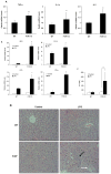
Figure 5. FXR has anti-inflammatory activity in vivo
(A) Quantitative real time PCR analysis of the expression of pro-inflammatory genes in livers from 12-month-old wild-type (WT) or FXR−/− (FXR KO) mice (n=4). *P < .05 compared to the WT group. (B) Quantitative real time PCR analysis of the expression of pro-inflammatory genes in livers from wild-type (WT) and FXR−/− (FXR KO) mice that were treated with a single dose of LPS (20 mg/kg) or PBS (as controls) (n=5). *P < .05 compared to the control group of FXR−/−.(C) ALT levels in wild-type (WT) and FXR−/− (FXR KO) mice that were treated with either vehicle (PBS) or 20 mg/kg LPS (n=5). *P < .05 compared to the control group of FXR−/−. (D) Representative H&E staining of liver sections from wild-type and FXR−/− livers (200×). Black arrow indicates infiltrated inflammatory cells; white arrow indicates regions of necrosis. (E) Representative TUNEL staining of sections from wild-type (WT) and FXR−/− livers (200×) and statistical analysis of the number of TUNEL-positive cells per total number of cells. The number of cells in at least 20 microscopic fields was counted. *P < .05 compared to the control group of FXR−/− (n=5). (F, G) Quantitative real-time PCR analysis of expression of FXR target genes and pro-inflammatory genes in livers from wild-type (F) or FXR−/− (G) mice that were transfected with adenovirus expressing VP16 alone (ConAd) or adenovirus expressing constitutively active murine FXRα2 (FXRAd) (n=4). The mice received a single dose of LPS (30 mg/kg) or PBS as control. *P < .05 compared to the corresponding control groups.
Figure 6
Figure 6. FXR activation suppressed NF-κB transcriptional activity by decreasing the binding between NF-κB and DNA sequences
(A) Immunoblot analysis for p65 and lamin B1 from nuclear protein pools in livers of wild-type or FXR−/− mice that were treated with a single dose of LPS (20 mg/kg) or PBS (as controls) (n=5). Lamin B1 was served as a loading control. Con, control; WT, wild-type; FXR KO, FXR knockout. (B) Immunoblot analysis for p65 and lamin B1 from nuclear protein pools in livers of wild-type mice that were transfected with adenovirus expressing VP16 alone (ConAd) or adenovirus expressing constitutively active murine FXRα2 (FXRAd) (n=4). The mice received a single dose of LPS (30 mg/kg) or PBS as control. Con, control. (C, D) HepG2 cell nuclear extracts were used in immunoblot analysis for p65 and lamin B1 (C) and a gel mobility shift assay with NF-κB binding site as a probe (D). HepG2 cells were transfected with p65 expression plasmid or control plasmid with or without co-transfection of FXR plasmid. After 24-h transfection, cells were treatment with 2μM GW4064 or DMSO (control) for 24 h. Finally, cells were collected and then nuclear proteins were extracted for immunoblot ananlysis or gel shift assay.
Figure 7
Figure 7. FXR activation has no effect on NF-κB anti-apoptotic target genes
(A) Quantitative real-time PCR analysis of the expression of Bfl-1, GADD45β, and cIAP1 in wild-type mouse primary hepatocytes that were pretreated with vehicle (DMSO), GW4064 (2 μM) or 6ECDCA (3 μM) for 18 h before treatment with TNF-α (10 ng/ml) for 6 h. (B, C) Quantitative real-time PCR analysis of the expression of Traf1, Traf2, cIAP2 and Bfl-1 in livers from wild-type (B) or FXR−/− (C) mice that were transfected with ConAd or FXRAd (n=4). The mice received a single dose of LPS (30 mg/kg) or PBS as control.
Figure 8
Figure 8. Activation of NF-κB by TPA, LPS and p65 overexpression antagonizes FXR transactivation
(A, B) Relative luciferase activities of HepG2 cells that were co-transfected with the FXR reporter plasmid EcRE-LUC, the control plasmid phRL-TK, and/or FXR/RXR expression plasmids, and pre-treated with GW4064 or vehicle (DMSO) for 18 h before treatment with TPA (50 nM) (A) or LPS (1 μg/mL) (B) for 6 hours. *P < .05, **P < .005 (n=3); (C) Relative luciferase activities of HepG2 cells were co-transfected with the FXR reporter plasmid EcRE-LUC, the control plasmid phRL-TK, and increasing amounts of a p65 expression plasmid at 0.5:1, 1:1 or 3:1 ratios with FXR/RXR expression plasmids, and then treated with GW4064 or vehicle (DMSO) for 24 h. *P < .05 compared to the control group; #P < .05 (n=3).

Comment in

References

    1. Gujral JS, Farhood A, Bajt ML, Jaeschke H. Neutrophils aggravate acute liver injury during obstructive cholestasis in bile duct-ligated mice. Hepatology. 2003;38:355–363. - PubMed
    1. Gu X, Ke S, Liu D, Sheng T, Thomas PE, Rabson AB, Gallo MA, et al. Role of NF-kappaB in regulation of PXR-mediated gene expression: a mechanism for the suppression of cytochrome P-450 3A4 by proinflammatory agents. J Biol Chem. 2006;281:17882–17889. - PubMed
    1. Karin M, Greten FR. NF-kappaB: linking inflammation and immunity to cancer development and progression. Nat Rev Immunol. 2005;5:749–759. - PubMed
    1. Pikarsky E, Porat RM, Stein I, Abramovitch R, Amit S, Kasem S, Gutkovich-Pyest E, et al. NF-kappaB functions as a tumour promoter in inflammation-associated cancer. Nature. 2004;431:461–466. - PubMed
    1. Elsharkawy AM, Mann DA. Nuclear factor-kappaB and the hepatic inflammation-fibrosis-cancer axis. Hepatology. 2007;46:590–597. - PubMed (VSports手机版)

V体育平台登录 - Publication types

MeSH terms

Substances