<style draggable="qxJVhB"><noframes date-time="9Tmhy7c"><code dropzone="f1EDFw"></code> V体育官网 - Skip to main page content
U.S. flag

An official website of the United States government

Dot gov

The . gov means it’s official. Federal government websites often end in . gov or VSports app下载. mil. Before sharing sensitive information, make sure you’re on a federal government site. .

Https

The site is secure V体育官网. The https:// ensures that you are connecting to the official website and that any information you provide is encrypted and transmitted securely. .

. 2008 Oct 10;322(5899):265-8.
doi: 10.1126/science.1162403.

Small molecule-induced allosteric activation of the Vibrio cholerae RTX cysteine protease domain

Affiliations

Small molecule-induced allosteric activation of the Vibrio cholerae RTX cysteine protease domain

Patrick J Lupardus et al. Science. .

Abstract

Vibrio cholerae RTX (repeats in toxin) is an actin-disrupting toxin that is autoprocessed by an internal cysteine protease domain (CPD). The RTX CPD is efficiently activated by the eukaryote-specific small molecule inositol hexakisphosphate (InsP6), and we present the 2. 1 angstrom structure of the RTX CPD in complex with InsP6. InsP6 binds to a conserved basic cleft that is distant from the protease active site VSports手机版. Biochemical and kinetic analyses of CPD mutants indicate that InsP6 binding induces an allosteric switch that leads to the autoprocessing and intracellular release of toxin-effector domains. .


V体育2025版 - Figures

Fig. 1
Fig. 1
InsP6 activates the V. cholerae RTX cysteine protease domain (CPD) and the architecture of InsP6-CPD complex. (A) Activation of RTX cysteine protease domain autocleavage by InsP6. Recombinant RTX CPD (amino acids 3391-3650) was incubated with the indicated concentrations of InsP6 for 2 h, and autocleavage was assessed by SDS-PAGE and Coomassie staining (representative gel inset). Reactions were performed in triplicate, and the amount of autocleaved protein relative to the total protein amount was analyzed by densitometry, averaged and plotted versus concentration of InsP6. Fifty-percent of wildtype CPD was autoprocessed (AC50) at 0.85 ± 0.02 nM InsP6 (mean ± SD). (B) SPR analysis of InsP6 binding to wildtype biotinylated CPD immobilized on a streptavidin-coupled surface. Representative sensorgram (inset) shows binding of InsP6 to the CPD-bound surface over a concentration of 0.1 to 100 μM. Equilibrium binding analysis indicates a dissociation constant (KD) of 1.3 ± 0.2 μM (SD). (C) Structure of the CPD-InsP6 complex viewed from above the InsP6-binding site. (D) A view of the structure rotated ~135° to show the active site. The InsP6 binding site and active site are separated by a 22 amino acid β-hairpin structure (labeled ‘β-flap’). InsP6 and the side chains of the catalytic dyad (Cys140/His91) are shown as stick models.
Fig. 2
Fig. 2
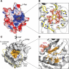
The InsP6 binding and active sites. (A) Electrostatic surface potential of the CPD as viewed from above the InsP6 binding site. Blue denotes positively charged surface; red denotes negatively charged surface. InsP6 is shown in the binding site as a stick model. (B) Close-up view of the InsP6 binding site. Side chains that directly interact with InsP6 are labeled and shown as yellow sticks. The electron density for InsP6 (2Fo-Fc) is contoured at 2σ. (C) Surface topology of the CPD active site. The P1 substrate pocket is highlighted in orange, and C140 and H91 in yellow and blue respectively. The N-terminus is shown as a yellow ribbon, terminating at Ile5 and highlighting the threading of this region along the surface of the core domain. The remaining residues not visible at the N-terminus are depicted as a yellow dashed line to illustrate approximate positioning of the chain during catalysis. (D) Close-up view of the P1 substrate pocket. Amino acids that line the pocket are labeled and colored orange. InsP6 is shown as in (B) to demonstrate the position of the catalytic site with respect to the InsP6 binding site.
Fig. 3
Fig. 3
β-flap mutations decouple CPD autocatalysis and RTX activity from InsP6 binding. (A) Comparison of autocleavage efficiency (AC50) versus InsP6 binding (KD) measured by SPR for mutations in the InsP6 binding site (left table) and β-flap (right tables, top and bottom). The β-flap region of the CPD is rainbow-colored starting with blue at the N-terminal end. β-flap, catalytic site, and visible InsP6-interacting side chains are shown as sticks. Data are expressed as mean ± SD. ND, not determinable. (B) Western blot analysis of RTX in supernatant harvested from log-phase V. cholerae cultures. Supernatants from V. cholerae strains harboring either an intact rtxA gene (wt), a null mutation in rtxArtxA), or point mutations in the region encoding the CPD domain of RTX (C140A is catalytic-dead; R182QK183N is mutated at two InsP6-binding residues; and W192A is a β-flap mutation) were blotted using an anti-CPD antibody. (C) Actin crosslinking induced upon incubation of V. cholerae with HFF cells. V. cholerae strains used in (A) were incubated with HFFs for 90 min, then HFF cells were lysed. Actin crosslinking was visualized by SDS-PAGE and Western blotting using an anti-actin antibody. The cross-linked forms of actin are labeled to the right.

V体育安卓版 - References

    1. Gordon VM, Leppla SH. Infect. Immun. 1994;62:333. - PMC - PubMed
    1. Reineke J, et al. Nature. 2007;446:415. - "VSports" PubMed
    1. Sheahan KL, Cordero CL, Satchell KJ. EMBO J. 2007;26:2552. - PMC - PubMed
    1. Satchell KJ. Infect. Immun. 2007;75:5079. - PMC - PubMed
    1. Cordero CL, Sozhamannan S, Satchell KJ. J. Clin. Microbiol. 2007;45:2289. - PMC (V体育平台登录) - PubMed

Publication types

"V体育平台登录" MeSH terms

V体育官网入口 - Associated data