Skip to main page content (VSports在线直播)
U.S. flag

An official website of the United States government

Dot gov

The . gov means it’s official. Federal government websites often end in . gov or . mil. Before sharing sensitive information, make sure you’re on a federal government site VSports app下载. .

Https

The site is secure V体育官网. The https:// ensures that you are connecting to the official website and that any information you provide is encrypted and transmitted securely. .

. 2008 Nov;9(11):1279-87.
doi: 10.1038/ni.1653. Epub 2008 Sep 21.

The immunity-related GTPase Irgm1 promotes the expansion of activated CD4+ T cell populations by preventing interferon-gamma-induced cell death (V体育ios版)

Affiliations

The immunity-related GTPase Irgm1 promotes the expansion of activated CD4+ T cell populations by preventing interferon-gamma-induced cell death (V体育2025版)

Carl G Feng et al. Nat Immunol. 2008 Nov.

"VSports app下载" Abstract

Mice deficient in the interferon-gamma (IFN-gamma)-inducible, immunity-related GTPase Irgm1 have defective host resistance to a variety of intracellular pathogens. This greater susceptibility to infection is associated with impaired IFN-gamma-dependent macrophage microbicidal activity in vitro. Here we show that Irgm1 also regulated the survival of mature effector CD4(+) T lymphocytes by protecting them from IFN-gamma-induced autophagic cell death VSports手机版. Mice deficient in both IFN-gamma and Irgm1 were 'rescued' from the lymphocyte depletion and greater mortality that occurs in mice singly deficient in Irgm1 after mycobacterial infection. Our studies identify a feedback mechanism in the T helper type 1 response that limits the detrimental effects of IFN-gamma on effector T lymphocyte survival while promoting the antimicrobial functions of IFN-gamma. .


Conflict of interest statement

The authors have no conflicting financial interests.

Figures

Figure 1
Figure 1
Irgm1–/– CD4+ T cell populations fail to expand following TCR engagement. 3H thymidine incorporation of (a) wild-type (WT) or Irgm1–/– CD4+ T cells activated with soluble anti-CD3 mAb and irradiated syngenic WT splenocytes, (b) OT-II or Irgm1–/– OT-II CD4+ T cells stimulated with OVA protein or peptide in the presence of syngenic splenic DC, was measured at 72 h. Kinetic analysis of (c)3H thymidine incorporation and (d) cell numbers in OVA(323-339) peptide stimulated OTII or Irgm1–/– OT-II splenic cultures. (e) Flow cytometric analysis of T cell activation and proliferation. OVA peptide stimulated OT-II or Irgm1–/– OT-II CD4+ T cells were pulsed with BrdU during the final 4 of 72 h culture and then co-stained with mAb to CD25 and BrdU. The numbers indicate the percentage of viable CD4+ cells. (f) IL-2 produced by OT-II (open bar) or Irgm1–/– OT-II (closed bar) CD4+ T cells stimulated with OVA peptide in the presence of splenic DC measured at 72 h by ELISA. Data shown are the mean concentration (± SD) of triplicate cultures. Results shown are the mean c. p. m. (a–c) or viable cell numbers (d) (± SD) of triplicate cultures. Data are representative of more than five (a, b) or three (c–f) independent experiments with similar results.
Figure 2
Figure 2
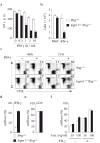
Irgm1 is essential for the expansion of CD4+ T cells in the presence of IFN-γ. (a) Immunoblot analysis of expression of Irgm1 in WT CD4+ T cells. FACS-sorted naive CD4+ lymphocytes were activated with immobilized agonistic CD3 (10 μg/ml) and CD28 (2 μg/ml) mAb in the presence or absence of IFN-γ neutralizing mAb for 48 h and lysates prepared. (b) Concentrations of IFN-γ produced by OT-II (open bars) or Irgm1–/– OT-II (closed bars) CD4+ T cells stimulated with OVA peptide in the presence of splenic DC under polarized or non-polarized (N) conditions in 72 h cultures were measured by ELISA. Data shown are the mean concentration (± SD) of triplicate cultures. ND: not detected. (c) Accumulation of polarized and non-polarized OT-II (open symbols) or Irgm1–/– OT-II (closed symbols) CD4+ T cells following peptide stimulation quantified by viable cell count. (d) Viable cell numbers of OT-II (open bars) or Irgm1–/– OT-II (closed bars) CD4+ T cells 72 h after restimulation with OVA peptide in the presence of T cell-depleted APCs. (e) Flow cytometric analysis of viability and division of OT-II or Irgm1–/– OT-II CD4+ T cells 72 h after secondary peptide stimulation. Peptide-stimulated OT-II or Irgm1–/– OT-II CD4+ T cells were rested in IL-2 for 7 days, and then CFSE-labeled and restimulated with OVA peptide in the presence of T cell-depleted APCs. (f) Flow cytometric analysis of expansion of CFSE-labeled OTII or Irgm1–/– OT-II CD4+ T cells activated with OVA peptide in the presence of absence of IFN-γ neutralizing mAb. (g)3H thymidine incorporation of WT, Irgm1–/–, Ifng–/–, Irgm1–/–Ifng–/– CD4+ T cells stimulated with soluble agonistic CD3 mAb and irradiated syngenic Ifng–/– splenocytes for 72 h. Results are the mean viable cell numbers (c, d) or c.p.m. (g) (± SD) of triplicate cultures. FACS analysis was performed after gating on CD4+ T cells and the numbers indicate the percentage of CD4+ cells (e, f). All data shown are representative of two independent experiments with similar results.
Figure 3
Figure 3
Irgm1 promotes cell survival during pathogen-driven TH1 responses and prevents IFN-γ-dependent mortality in mycobacteria-infected mice. (a) Survival of CD4+ T cells in vivo. CD45.2+Ifng–/– (open symbols) or Irgm1–/–Ifng–/– (closed symbols) CD4+ lymphocytes pre-activated with agonistic CD3 mAb in vitro were transferred i.p. into congenic CD45.1+ C57BL/6.SJL recipients that were left untreated or inoculated i.p. with T. gondii or M. avium 3 days earlier. Percentage of donor CD4+ T cells in PEC collected at day 4 after transfer was determined by flow cytometry. (b) Survival of M. avium infected WT or KO mice (n = 5 per genotype) was monitored through the course of infection. (c) Splenic bacterial loads, (d) circulating and (e) splenic lymphocyte numbers were determined at wk4 after M. avium infection (n = 5). Data shown are representative of two independent experiments with each symbol representing an individual mouse.
Figure 4
Figure 4
IFN-γ directly induces death of Irgm1–/– CD4+ T cells. (a)3H thymidine incorporation. Naive CD4+ lymphocytes were stimulated with agonistic CD3 mAb and IFN-γ–deficient irradiated APCs for 72 h. IL-2 (10 U/ml)) was added 24 h after initiation of the cultures. Data shown are the mean c.p.m. (± SD) of triplicate cultures. (b) Accumulation of CD4+ T cells in the presence of IFN-γ (5 U/ml) at 72 h. Data shown are the mean viable cell numbers (± SD) of triplicate cultures. (c) Flow cytometric analysis of cell viability and division. CD4+ T cells were CFSE-labeled and activated as in (a) with T-depleted Ifng–/– APCs in the presence or absence of IFN-γ (5 U/ml). The numbers shown are the percentage of CFSE+CD4+ T cell populations in the cultures. (d) IFN-γ (e) agonistic CD3 mAb and (f) FasL triggered death of Ifng–/– or Irgm1–/–Ifng–/– T cells that were previously activated with anti-CD3 mAb and expanded in IL-2 medium. The cell loss was determined 48 h after each treatment with PI staining. In a, b, d, e and f, open bars: Ifng–/–, closed bars: Irgm1–/–Ifng–/–. Data are representative of three independent experiments with similar results.
Figure 5
Figure 5
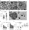
Irgm1 prevents IFN-γ induced death of CD4+ T cells by regulating autophagy. EM analysis of (a)Ifng–/– or (b - f)Irgm1–/–Ifng–/– CD4+ lymphocytes that have been exposed to IFN-γ (5 U/ml) for 30 h. Original magnification: ×1000. (c) A magnified view of the KO cell boxed in (b) with two autophagic vacuoles indicated with arrows heads; (d) the numbers of the vacuoles quantified blindly on 50 randomly selected EM sections with more than 400 cells. Each symbol represents one section; (e) a representative image of Irgm1–/– CD4+ T cells with numerous vacuoles containing dense materials and (f) two Irgm1–/– CD4+ cells with vacuolated cytoplasma. (g) IFN-γ-induced death of CD4+ T cells in the presence or absence of PI3K-inhibitors Wortamanin or Ly294002. Data are representative of three independent experiments. (h) IFN-γ induced death of Ifng–/– or Irgm1–/–Ifng–/– CD4+ T cells transfected with control or Beclin1 specific siRNA. Reduction in expression of the Beclin1 protein was measured by western blot. The data shown are means (± SD) of triplicate cultures and are representative of three independent experiments. Cell death (g, h) was determined 48 h after IFN-γ exposure by flow cytometry with PI staining.

Comment in

References

    1. Gett AV, Sallusto F, Lanzavecchia A, Geginat J. T cell fitness determined by signal strength. Nat Immunol. 2003;4:355–60. - PubMed
    1. Khoruts A, Mondino A, Pape KA, Reiner SL, Jenkins MK. A natural immunological adjuvant enhances T cell clonal expansion through a CD28-dependent, interleukin (IL)-2-independent mechanism. J Exp Med. 1998;187:225–36. - PMC - PubMed
    1. Croft M. Co-stimulatory members of the TNFR family: keys to effective T-cell immunity? Nat Rev Immunol. 2003;3:609–20. - VSports - PubMed
    1. Krammer PH, Arnold R, Lavrik IN. Life and death in peripheral T cells. Nat Rev Immunol. 2007;7:532–42. - PubMed
    1. Green DR. Fas Bim boom! Immunity. 2008;28:141–3. - PubMed

Publication types (V体育官网入口)

MeSH terms