<acronym id="DyjkU"></acronym>
"V体育安卓版" Skip to main page content
U.S. flag

An official website of the United States government

Dot gov

The . gov means it’s official. Federal government websites often end in VSports app下载. gov or . mil. Before sharing sensitive information, make sure you’re on a federal government site. .

Https

The site is secure. The https:// ensures that you are connecting to the official website and that any information you provide is encrypted and transmitted securely. V体育官网.

. 2008 Oct 1;181(7):4666-75.
doi: 10.4049/jimmunol.181.7.4666.

Proinflammatory S100 proteins regulate the accumulation of myeloid-derived suppressor cells

Affiliations

Proinflammatory S100 proteins regulate the accumulation of myeloid-derived suppressor cells

Pratima Sinha et al. J Immunol. .

VSports - Abstract

Chronic inflammation is a complex process that promotes carcinogenesis and tumor progression; however, the mechanisms by which specific inflammatory mediators contribute to tumor growth remain unclear. We and others recently demonstrated that the inflammatory mediators IL-1beta, IL-6, and PGE(2) induce accumulation of myeloid-derived suppressor cells (MDSC) in tumor-bearing individuals. MDSC impair tumor immunity and thereby facilitate carcinogenesis and tumor progression by inhibiting T and NK cell activation, and by polarizing immunity toward a tumor-promoting type 2 phenotype. We now show that this population of immature myeloid cells induced by a given tumor share a common phenotype regardless of their in vivo location (bone marrow, spleen, blood, or tumor site), and that Gr1(high)CD11b(high)F4/80(-)CD80(+)IL4Ralpha(+/-)Arginase(+) MDSC are induced by the proinflammatory proteins S100A8/A9. S100A8/A9 proteins bind to carboxylated N-glycans expressed on the receptor for advanced glycation end-products and other cell surface glycoprotein receptors on MDSC, signal through the NF-kappaB pathway, and promote MDSC migration. MDSC also synthesize and secrete S100A8/A9 proteins that accumulate in the serum of tumor-bearing mice, and in vivo blocking of S100A8/A9 binding to MDSC using an anti-carboxylated glycan Ab reduces MDSC levels in blood and secondary lymphoid organs in mice with metastatic disease. Therefore, the S100 family of inflammatory mediators serves as an autocrine feedback loop that sustains accumulation of MDSC. Since S100A8/A9 activation of MDSC is through the NF-kappaB signaling pathway, drugs that target this pathway may reduce MDSC levels and be useful therapeutic agents in conjunction with active immunotherapy in cancer patients VSports手机版. .


Conflict of interest statement

Disclosures

The authors have no financial conflict of interest.

Figures

FIGURE 1
FIGURE 1
MDSC present in the bone marrow, blood, spleen, or lungs of BALB/c mice with the 4T1 mammary carcinoma are Gr1highCD11bhigh F4/80IL-4Rα+/− CD80+Arginase+and have a mixed nuclear phenotype. A, Spleens, blood, bone marrow, and metastatic lung from BALB/c mice with 33-day primary tumors (11.5 mm in diameter) were harvested and the cells were stained with Abs to Gr1, CD11b, F4/80, IL-4Rα, CD80, arginase, iNOS, Ly-6C, and Ly-6G. Gr1+CD11b+ populations were gated and further analyzed by flow cytometry for F4/80, IL-4Rα, CD80, arginase, or iNOS. B, CD11b+ cells of A were gated and analyzed for Ly6G and Ly6C. C, Cell populations from A were stained with Diff-Quik and analyzed by microscopy. Each panel shows representative cells assembled from three to six fields per MDSC sample. D, Purified Gr1+CD11b+ cells from the spleen, bone marrow, blood, or metastatic lungs were cocultured with transgenic CD4+ D011.10 or CD8+ Clone 4 splenocytes plus OVA323–339 or HA518–526 peptides, respectively, and T cell activation measured by tritiated thymidine uptake.
FIGURE 1
FIGURE 1
MDSC present in the bone marrow, blood, spleen, or lungs of BALB/c mice with the 4T1 mammary carcinoma are Gr1highCD11bhigh F4/80IL-4Rα+/− CD80+Arginase+and have a mixed nuclear phenotype. A, Spleens, blood, bone marrow, and metastatic lung from BALB/c mice with 33-day primary tumors (11.5 mm in diameter) were harvested and the cells were stained with Abs to Gr1, CD11b, F4/80, IL-4Rα, CD80, arginase, iNOS, Ly-6C, and Ly-6G. Gr1+CD11b+ populations were gated and further analyzed by flow cytometry for F4/80, IL-4Rα, CD80, arginase, or iNOS. B, CD11b+ cells of A were gated and analyzed for Ly6G and Ly6C. C, Cell populations from A were stained with Diff-Quik and analyzed by microscopy. Each panel shows representative cells assembled from three to six fields per MDSC sample. D, Purified Gr1+CD11b+ cells from the spleen, bone marrow, blood, or metastatic lungs were cocultured with transgenic CD4+ D011.10 or CD8+ Clone 4 splenocytes plus OVA323–339 or HA518–526 peptides, respectively, and T cell activation measured by tritiated thymidine uptake.
FIGURE 2
FIGURE 2
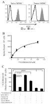
MDSC have glycoprotein receptors for and bind S100A8/A9 proteins. A, Leukocytes from the blood of tumor-free or 4T1 tumor-bearing BALB/c mice were stained with Gr1, CD11b, and GB3.1 mAbs. Gr1+CD11b+ cells were gated and analyzed for expression of GB3.1 receptors. B, Cell membranes of MDSC derived from 4T1 tumor-bearing mice were incubated with increasing concentrations of 125I-S100A8/A9 proteins for 1 h at 4°C followed by washing, and membrane bound radioactivity was measured. Saturation binding kinetic analysis was performed using GraphPad Prism. Values represent mean ± SD of two independent experiments. C, Inhibition of binding of 125I-S100A8/A9 to MDSC. MDSC membranes were incubated with 125I-S100A8/A9 proteins (20 nM) in the presence or absence of mAbGB3.1, anti-RAGE, anti-S100A8 (10-fold molar excess), cold ligand (100-fold molar excess), or isotype control Abs. Membrane-bound radioactivity was determined as in A. Data are from one of two independent experiments.
FIGURE 3
FIGURE 3
MSDC express and secrete S100A8/A9, and migrate and signal through NF-κB in response to S100A8/A9. A, Circulating white blood cells pooled from five 4T1 tumor-bearing mice (left panel) or pooled white blood cells purified by MACS sorting for Gr-1 from 60 tumor-free mice (right panel) were stained with fluorescent Abs to Gr1 and CD11b and analyzed by flow cytometry. B, MDSC contain elevated levels of mRNA for proinflammatory mediators. Real-time RT-PCR was performed on mRNA obtained from the purified cells of A. Expression levels are presented as the number of threshold cycles ± SD needed to detect a product. Fold increase is the relative increase in MDSC from tumor-bearing mice vs tumor-free mice. Values for the HG are the average of five genes: GAPDH, Hprt1, Hsp90ab1, actin β cytoplasmic, and glucuronidase β. CXCL1 is included as an example of a poorly expressed gene. C, Lysates of the Gr1+CD11b+ cells shown in A were electrophoresed on 12% SDS-PAGE gels, and Western blotted with Abs to S100A8, S100A9, HMGB1, and RAGE. D, MDSC purified from BALB/c 4T1 tumor-bearing or from tumor-free mice were cultured for 16 h, and the supernatants were assayed by ELISA for S100A8/A9 proteins. Data are from one of two independent experiments. E, NF-κB is phosphorylated in MDSC following binding of S100A8/A9 proteins. MDSC from tumor-bearing or tumor-free mice were cocultured with S100A8/A9 or TNF-α for 10 min and cell lysates were screened for phosphorylated-NF-κB p65 (ser536) by ELISA. Data are from one of two independent experiments. F, CM from 4T1 tumor cells was tested by ELISA for S100A8/A9. G, 4T1 tumor cells secrete S100A8/A9 proteins that are chemotactic for MDSC. Blood MDSC (>90% Gr1+CD11b+ cells) were tested by chemotaxis assay for their migration in response to 4T1 CM. Data are pooled from two independent experiments.
FIGURE 4
FIGURE 4
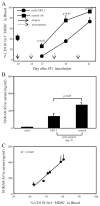
mAbGB3.1 reduces MDSC and S100A8/A9 proteins in the blood of tumor-bearing mice. A, mAbGB3.1 treatment reduces serum levels of MDSC in tumor-bearing mice. BALB/c mice were inoculated on day 0 with 4T1 tumor cells, their primary tumors were removed on day 20, and GB3.1 or control Ab treatment was started 3 days later (day 24) and continued once weekly. Mice were bled 72 h after each Ab treatment and their white blood cells stained for Gr1 and CD11b. At the time of surgery, primary tumor diameters were 5.09 ± 0.76 and 5.36 ± 0.68 mm, and percent Gr1+CD11b+ MDSC in the blood were 41.25 ± 3.98 and 44.65 ± 6.60%, for the mAbGB3.1 and control Ab-treated groups, respectively. Data are the average ± SD of four mice in each group. Experiment was terminated on day 42 when mice were moribund. Data are from one of two independent experiments. B, mAbGB3.1 treatment reduces serum levels of S100A8/A9 proteins. GB3.1 and control Ab-treated postsurgery mice from A were bled on day 41 (20 days after surgery), and the serum assayed by ELISA for S100A8/A9 proteins. Serum from tumor-free (naive) mice was included for comparison. C, Serum S100A8/A9 levels are proportional to the amount of circulating MDSC. Tumor-bearing mice were bled and serum levels of S100A8/A9 were determined by ELISA and the percent of MDSC determined by flow cytometry. Data are from one of two independent experiments.
FIGURE 5
FIGURE 5
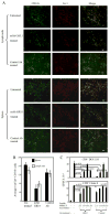
Blocking S100A8/A9 binding reduces MDSC accumulation in vivo in tumor-bearing mice but does not alter immunosuppressive activity of MDSC on a per cell basis. A, Spleen and lymph nodes from BALB/c mice with 41 day 4T1-tumors were cryo-preserved and stained for CD11b and Gr-1. Representative sections are shown. Scale bars indicate 100 microns. B, Average number of Gr1+CD11b+ cells counted in three high power fields of lymph nodes and spleen for untreated, mAb GB3.1-treated, and control Ab-treated groups. C, CD4+ DO11.10 or CD8+ Clone 4 transgenic splenocytes were cocultured with OVA or HA peptide, respectively, in the presence or absence of graded doses of MACS-sorted splenic Gr1+ CD11b+ MDSC (>90% Gr1+CD11b+) from mAbGB3.1 or control mAb-treated postsurgery BALB/c mice. BALB/c mice were inoculated on day 0 with 4T1 cells, primary tumors were removed on day 26 (primary tumor diameters were 4.64 ± 0.55 and 5.33 ± 0.32 for control and mAbGB3.1 depletion groups, respectively), mAbGB3.1 or control Ab treatment was started on day 29, and MDSC were harvested 1 day after the last Ab treatment on day 45. T cell activation was measured by [3H]thymidine incorporation. Data are from one of two independent experiments.

References

    1. Balkwill F, Mantovani A. Inflammation and cancer: back to Virchow? Lancet. 2001;357:539–545. - PubMed
    1. Coussens LM, Werb Z. Inflammation and cancer. Nature. 2002;420:860–867. - PMC - PubMed
    1. Ruegg C. Leukocytes, inflammation, and angiogenesis in cancer: fatal attractions. J Leukocyte Biol. 2006;80:682–684. - PubMed
    1. Luo JL, Maeda S, Hsu LC, Yagita H, Karin M. Inhibition of NF-κB in cancer cells converts inflammation- induced tumor growth mediated by TNFα to TRAIL-mediated tumor regression. Cancer Cell. 2004;6:297–305. - PubMed
    1. Karin M, Greten FR. NF-κB: linking inflammation and immunity to cancer development and progression. Nat Rev Immunol. 2005;5:749–759. - VSports手机版 - PubMed

V体育官网 - Publication types

"V体育2025版" MeSH terms