"V体育官网入口" Skip to main page content
U.S. flag

An official website of the United States government

Dot gov

The . gov means it’s official. Federal government websites often end in . gov or . mil VSports app下载. Before sharing sensitive information, make sure you’re on a federal government site. .

Https

The site is secure V体育官网. The https:// ensures that you are connecting to the official website and that any information you provide is encrypted and transmitted securely. .

. 2008 Aug 15;29(2):272-82.
doi: 10.1016/j.immuni.2008.05.016. Epub 2008 Jul 24.

"V体育平台登录" Toll-like receptors activate innate and adaptive immunity by using dendritic cell-intrinsic and -extrinsic mechanisms

Affiliations

"VSports最新版本" Toll-like receptors activate innate and adaptive immunity by using dendritic cell-intrinsic and -extrinsic mechanisms

VSports最新版本 - Baidong Hou et al. Immunity. .

Abstract

Toll-like receptors (TLRs) play prominent roles in initiating immune responses to infection, but their roles in particular cell types in vivo are not established. Here we report the generation of mice selectively lacking the crucial TLR-signaling adaptor MyD88 in dendritic cells (DCs) VSports手机版. In these mice, the early production of inflammatory cytokines, especially IL-12, was substantially reduced after TLR stimulation. Whereas the innate interferon-gamma response of natural killer cells and of natural killer T cells and the Th1 polarization of antigen-specific CD4(+) T cells were severely compromised after treatment with a soluble TLR9 ligand, they were largely intact after administration of an aggregated TLR9 ligand. These results demonstrate that the physical form of a TLR ligand affects which cells can respond to it and that DCs and other innate immune cells can respond via TLRs and collaborate in promoting Th1 adaptive immune responses to an aggregated stimulus. .


"VSports注册入口" Conflict of interest statement

COMPETING INTERESTS STATEMENT

The authors declare they have no competing financial interests.

Figures

Figure 1
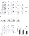
Figure 1. Defective inflammatory cytokine responses in the DC-MyD88 KO mice
(A and B) IL-12 expression in control (Flox) or DC-MyD88 KO (Flox/Cre) mice after i.v. injection with PBS or CpG. (A) Splenocytes isolated one hour after injection were stained for intracellular IL-12p40. Shown are representative FACS plots of gated CD11chi DCs for one of six mice per group analyzed on two separated days. (B) Amounts of IL-12p70 in the serum of mice, as measured by ELISA, at two hours after i.v. injection of CpG (n=5 for Flox/Cre mice). Amounts of IL-12p70 in PBS-treated mice all were under detection limit (not shown). (C) Induction of IL-12p40 mRNA in the spleen at one hour after i.v. injection of Pam3CSK4 (TLR1/2), LPS (TLR4), flagellin (TLR5), CpG (TLR9), or i.p. injection of Imiquimod (TLR7). (D) Induction of inflammatory cytokine mRNA in the spleen at one hour after i.v. injection of CpG. “fold-induction” (mean+s.d. of 3 mice) is relative to the abundance in vehicle-treated mice. Numbers on top of the bars are the fold difference between the induction levels in the control and the DC-MyD88 KO mice. Statistical comparison is between the DC-MyD88 KO mice and the control mice. *, P<0.05; **, P<0.01. Data are representative of two separate experiments.
Figure 2
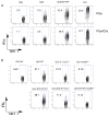
Figure 2. The physical form of CpG influences its interaction with immune cells
(A) WT mice were injected i.v. with CpG, Cy5.5-labeled CpG or Cy5.5-labeled CpG complexed with DOTAP. After 30 minutes, up-take of CpG was examined by flow cytometry on gated conventional DCs (cDC; CD11chi, I-Ab+), plasmacytoid DCs (pDC; CD11cint, B220+, Ly6C+, CD19), macrophages (Mφ, CD11bint, F4/80hi, Ly6G, SSChi), monocytes (Mo, CD11b+, F4/80+, Ly6C+, CD11c, Ly6G, SSClow) or NK cells (NK1.1+, TCRβ) in the spleen. Shown are representative FACS plots for one of two mice per group. (B) Splenic CD11chi DCs of WT mice were stained for intracellular IL-12p40 one hour after injection with the indicated TLR ligands. Shown are representative FACS plots for one of four mice per group analyzed on two separate days. (C) Control (Flox) or DC-MyD88 KO (Flox/Cre) mice were injected i.v. with indicated TLR ligands. After one hour, splenocytes were stained for intracellular TNFα. Shown are representative contour plots of gated CD11b+, F4/80+, Ly6C+, SSClow, CD11c, NK1.1, B220 cells from one of four mice per group analyzed on two separate days. (D) Induction of inflammatory cytokine mRNA in the spleen at one hour after injection i.v. with CpG complexed with DOTAP. “fold-induction” and fold difference were calculated as in Figure 1. Statistical comparison is between the DC-MyD88 KO mice and the control mice. *, P<0.05; **, P<0.01. Data are representative of two separate experiments.
Figure 3
Figure 3. Role of MyD88 in DCs for the IFNγ response of NK cells to TLR stimulation in vivo
(A and B) Mice were injected with the indicated TLR ligands. Five hours later, synthesis of IFNγ by splenic NK cells (NK1.1+, TCRβ) was assessed by intracellular staining and flow cytometry. (A) Representative contour plots of control (Flox) or DC-MyD88 KO (Flox/Cre) mice from one of six mice per group analyzed on two separate days. (B) Representative contour plot of WT (C57BL/6), IL-12p35−/−, Ifnar−/− and myd88−/− mice from one of four mice per group analyzed on two separate days.
Figure 4
Figure 4. Defects in DC maturation in the DC-MyD88 KO mice
(A) Control (Flox) or DC-MyD88 KO (Flox/Cre) mice were injected i.v. with CpG or CpG/DOTAP. After 12 hours, expression of CD86 (left), CD40 (middle), or I-Ab (right) on the surface of CD11chi DCs were assessed by flow cytometry. “Fold-induction” (mean+s.d. of four mice) of median fluorescence intensity (MFI) is relative to the expression levels in vehicle-treated mice. Statistical comparison is between the DC-MyD88 KO mice and the control mice. *, P<0.05; **, P<0.01. (B) Representative histograms of CD86 (left), CD40 (middle), or I-Ab on gated CD11chi DCs from control (WT or Flox), myd88−/− or DC-MyD88 KO (Flox/Cre) mice 12 hours after i.v. injection of CpG or CpG/DOTAP.
Figure 5
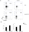
Figure 5. Clonal expansion and development of TH1 effector T cells in the DC-MyD88 KO mice
(A) Purified CD4+ T cells from OT-II transgenic mice with a congenic marker (Thy1.1+) were labeled with CSFE and adoptively transferred into Thy1.2+DC-MyD88 KO and control mice. One day later (day 0), the mice were immunized with OVA mixed with CpG (upper panels) or OVA mixed with CpG/DOTAP (lower panels). On days 4 or 7, lymphocytes from the spleen were harvested, and re-stimulated in vitro with PMA and ionomycin for 4 hours. The proliferation of OT-II T cells (identified as CD4+, B220, Thy1.1+) was tracked by CFSE dilution, and TH1 effector function development was assessed by intracellular IFNγ staining and flow cytometry. Shown are representative dot-plots of intracellular IFNγ staining and CFSE dilution on day 4 of single mice immunized with OVA + CpG or OVA + CpG/DOTAP. Numbers in the gates indicate percentage of OT-II T cells that were IFNγ+. (B) Absolute numbers of OT-II T cells (left panel) and IFNγ+ OT-II T cells (right panel) in the spleen at day 4 after immunization with OVA + CpG or OVA + CpG/DOTAP and in vitro restimulation, as calculated from the counts of total splenocytes and the percentages of OT-II T cells and IFNγ+ OT-II T cells in the spleens. Data are expressed as mean+s.d. of four mice, and are representative of three separate experiments. Statistical comparison is between the DC-MyD88 KO mice and the control mice. *, P<0.05.
Figure 6
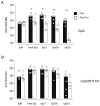
Figure 6. Antibody response in the DC-MyD88 KO mice
Control (Flox) and DC-MyD88 KO (Flox/Cre) mice were immunized i.p. with OVA together with CpG (upper panel) or CpG/DOTAP (lower panel) and were bled 7 and 14 days after immunization. Sera were serially diluted and OVA-specific antibody isotypes (IgM on day7 and total IgG, IgG1, IgG2b and IgG2c on day 14) were measured by ELISA. Data shown are the end-point titers of all samples. Statistical significance was calculated with Mann-Whitney U-test. *, P<0.05 **, P<0.01. Similar results were obtained in three (CpG) and two (CpG/DOTAP) separate experiments.

"V体育平台登录" Comment in

  • 2008: A MyD88 O, DC.
    Pulendran B, Cao W. Pulendran B, et al. Immunity. 2008 Aug 15;29(2):173-5. doi: 10.1016/j.immuni.2008.07.006. Immunity. 2008. PMID: 18701079

VSports注册入口 - References

    1. Adachi O, Kawai T, Takeda K, Matsumoto M, Tsutsui H, Sakagami M, Nakanishi K, Akira S. Targeted disruption of the MyD88 gene results in loss of IL-1- and IL-18-mediated function. Immunity. 1998;9:143–150. - PubMed
    1. Afkarian M, Sedy JR, Yang J, Jacobson NG, Cereb N, Yang SY, Murphy TL, Murphy KM. T-bet is a STAT1-induced regulator of IL-12R expression in naive CD4+ T cells. Nat Immunol. 2002;3:549–557. - PubMed
    1. Aida Y, Pabst MJ. Removal of endotoxin from protein solutions by phase separation using Triton X-114. J Immunol Methods. 1990;132:191–195. - PubMed
    1. Akira S. TLR signaling. Curr Top Microbiol Immunol. 2006;311:1–16. - PubMed
    1. Akira S, Uematsu S, Takeuchi O. Pathogen recognition and innate immunity. Cell. 2006;124:783–801. - PubMed

Publication types

"V体育平台登录" MeSH terms

Substances