<dfn draggable="ItPOtj"><del draggable="RV2Nw"><del lang="C4VS9"></del></del></dfn><sup date-time="kaFfE"></sup> "VSports最新版本" Skip to main page content
U.S. flag

An official website of the United States government

Dot gov

The . gov means it’s official. Federal government websites often end in . gov or VSports app下载. mil. Before sharing sensitive information, make sure you’re on a federal government site. .

Https

The site is secure V体育官网. The https:// ensures that you are connecting to the official website and that any information you provide is encrypted and transmitted securely. .

. 2008 Aug 8;321(5890):839-43.
doi: 10.1126/science.1156121. Epub 2008 Jul 24.

Human CHN1 mutations hyperactivate alpha2-chimaerin and cause Duane's retraction syndrome (VSports最新版本)

Affiliations

Human CHN1 mutations hyperactivate alpha2-chimaerin and cause Duane's retraction syndrome

Noriko Miyake et al. Science. .

"VSports" Abstract

Duane's retraction syndrome (DRS) is a complex congenital eye movement disorder caused by aberrant innervation of the extraocular muscles by axons of brainstem motor neurons. Studying families with a variant form of the disorder (DURS2-DRS), we have identified causative heterozygous missense mutations in CHN1, a gene on chromosome 2q31 that encodes alpha2-chimaerin, a Rac guanosine triphosphatase-activating protein (RacGAP) signaling protein previously implicated in the pathfinding of corticospinal axons in mice VSports手机版. We found that these are gain-of-function mutations that increase alpha2-chimaerin RacGAP activity in vitro. Several of the mutations appeared to enhance alpha2-chimaerin translocation to the cell membrane or enhance its ability to self-associate. Expression of mutant alpha2-chimaerin constructs in chick embryos resulted in failure of oculomotor axons to innervate their target extraocular muscles. We conclude that alpha2-chimaerin has a critical developmental function in ocular motor axon pathfinding. .


Figures

Figure 1
Figure 1. Duane’s retraction syndrome (DRS) and corresponding CHN1 mutations
(A) Affected member of pedigree JH with limited outward gaze (abduction) and narrowing of the palpebral fissure on attempted inward gaze (adduction) most obvious on leftward gaze. He also has bilateral exotropia on downgaze. (B) The seven DURS2-DRS pedigrees and corresponding heterozygous CHN1 mutations. (C) Schematic representation of α1- (top, 334 amino acids) and α2-chimaerin (bottom, 459 amino acids) protein. The isoforms contain identical C1 and RacGAP domains, while only α2-chimaerin contains an SH2 domain. Mutations alter residues unique to α2-chimaerin or common to both proteins, as indicated by the arrows. No mutations were found in the α1-chimaerin N-terminal sequence (highlighted in black).
Figure 2
Figure 2. Human developmental expression profile of α2-chimaerin mRNA by in situ hybridization
(A) Transverse section of CS15 human embryo showing α2-chimaerin mRNA expression (purple deposit) in midbrain (mb), hindbrain (rhombomere [rh] 2 indicated), spinal cord (sc). (B) Higher magnification of (A) showing expression in the ventricular layer of rh3 and rh4. (C) At CS16 expression is also seen in mb, hindbrain (hb), sc, and vestibulocochlear (viii) and vagus (x) nuclei. Higher magnifications of (C) show (D) expression in developing oculomotor neurons (o) and (E) in neurons of rh5 (developing abducens neurons) and rh6. In CS19 sagittal section (F), expression has declined in basal mb and hb and is now found in dorsal root ganglia (drg), cerebellum (cb), diencephalons (dc), and telencephalon (t). At later stages (G), expression is located in specific regions of the cortical plate (c), intermediate (i) and ventricular zone (v) of the forebrain (11 wpo). No signal was detected in corresponding sections hybridized with sense probe (fig. S4). Scale bars 1000μm (A, C), 100μm (B, E), 200μm (D), 2000μm (F), 500μm (G).
Figure 3
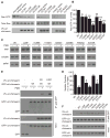
Figure 3. DURS2-DRS mutations enhance α2-chimaerin function in vitro.
(A) Rac-GTP levels were measured in HEK293T cells transfected with plasmids encoding for myc-ephexin, V5-empty vector, V5-α2-chimaerin wild-type, or V5-α2-chimaerin mutant. Rac-GTP levels are reduced by overexpression of wild-type α2-chimaerin compared to empty vector, and further reduced in cells expressing each mutant, while elevated with overexpression of a GEF, myc-ephexin (27). (B) Densitometric analysis of Rac-GTP levels normalized to total Rac and V5-α2-chimaerin levels. Values are expressed as percent of wild-type α2-chimaerin (mean+SEM, n = 6–10). The difference between the reduction of Rac-GTP levels for each mutant compared to wild-type α2-chimaerin is significant by one-way ANOVA with Dunnett’s adjustment (F=9.89, *p<0.03, **p<0.005, ***p<0.0001). (C) α2-chimaerin translocation examined by immunoblots of total, soluble and pellet fraction of wild-type and mutant α2-chimaerin +/- 10 PMA stimulation. (D) Graphical representation of translocation following PMA compared to pretreatment expressed as the percent of α2-chimaerin remaining in the soluble fraction (mean+SEM, n = 3). Enhanced translocation compared to wild-type is significant for L20F, Y143H, A223V, and P252Q by one-way ANOVA with Dunnett’s adjustment (F=21.00, *p<0.0001). (E) GFP-α2-chimaerin immunoprecipitates with V5-wild-type- or V5-L20F-α2-chimaerin in the presence of PMA, and minimally in its absence. (F) In the presence of PMA, immunoprecipitation of wild-type α2-chimaerin is enhanced by all mutant-α2-chimaerins compared to wild-type except G228S and E313K, which were equivalent to wild-type α2-chimaerin. Results were consistent over at least four independent experiments (also see fig. S6F&G).
Figure 4
Figure 4. α2-chimaerin overexpression results in stalling of developing chick oculomotor nerves
(A) Transverse section through E4 whole chick embryo showing wide neuroepithelial expression of α2-chimaerin mRNA including the hindbrain (hb), forebrain (fb), and trigeminal ganglion (tg). (B) Transverse section through E5-6 chick midbrain showing α2-chimaerin mRNA expression in the oculomotor nuclei (left nucleus circled in white). (C) Tabulated results of electroporated constructs. (D-I) Confocal image montages (white hatches denote image breaks) at E6 of electroporated oculomotor nerves (green) and extraocular muscles (red) labeled with anti-myosin antibody (D,E,G-I) or α-bungarotoxin (F); constructs as labeled. All GFP-control (D), 28% of wild-type (E), and only 5–13% of mutant α2-chimaerin electroporated oculomotor nerves extend normally from the midbrain neuroepithelium, at left, past the dorsal rectus muscle (DR), ciliary ganglion (*), and ventral (VR) and medial (MR) recti to innervate the first target, the ventral oblique (VO) muscle. Nerves expressing mutant α2-chimaerin have a higher incidence of aberrant branching (arrow in F with higher magnification inset) and defasciculation than wildtype (fig. S7). Most remarkably, 72% of wildtype (G), 87% of L20F (H), and 71% of G228S-α2-chimaerin (I) electroporated nerves stall in the vicinity of the DR muscle. Scale bars are 200 μm.

References

    1. Supplemental figures and materials and methods are available as supporting material on Science Online.

    1. Engle EC. Archives of Neurology. 2007 May;64:633. - PubMed
    1. Jen J, et al. Neurology. 2002 Aug 13;59:432. - PubMed (VSports app下载)
    1. Hotchkiss MG, Miller NR, Clark AW, Green WG. Archives of Ophthalmology. 1980 May;98:870. - PubMed
    1. Miller NR, Kiel SM, Green WR, Clark AW. Archives of Ophthalmology. 1982 Sep;100:1468. - V体育官网入口 - PubMed

Publication types

"VSports最新版本" MeSH terms

Associated data

LinkOut - more resources