"VSports在线直播" Skip to main page content
U.S. flag

An official website of the United States government

Dot gov

The . gov means it’s official VSports app下载. Federal government websites often end in . gov or . mil. Before sharing sensitive information, make sure you’re on a federal government site. .

Https

The site is secure. The https:// ensures that you are connecting to the official website and that any information you provide is encrypted and transmitted securely V体育官网. .

. 2008 Jul 29;105(30):10483-8.
doi: 10.1073/pnas.0804453105. Epub 2008 Jul 18.

V体育2025版 - Crystal structure of the complex between programmed death-1 (PD-1) and its ligand PD-L2

Affiliations

Crystal structure of the complex between programmed death-1 (PD-1) and its ligand PD-L2

Eszter Lázár-Molnár et al. Proc Natl Acad Sci U S A. .

VSports在线直播 - Abstract

Programmed death-1 (PD-1) is a member of the CD28/B7 superfamily that delivers negative signals upon interaction with its two ligands, PD-L1 or PD-L2. The high-resolution crystal structure of the complex formed by the complete ectodomains of murine PD-1 and PD-L2 revealed a 1:1 receptor:ligand stoichiometry and displayed a binding interface and overall molecular organization distinct from that observed in the CTLA-4/B7 inhibitory complexes. Furthermore, our structure also provides insights into the association between PD-1 and PD-L1 and highlights differences in the interfaces formed by the two PD-1 ligands (PD-Ls) Mutagenesis studies confirmed the details of the proposed PD-1/PD-L binding interfaces and allowed for the design of a mutant PD-1 receptor with enhanced affinity. These studies define spatial and organizational constraints that control the localization and signaling of PD-1/PD-L complexes within the immunological synapse and provide a basis for manipulating the PD-1 pathways for immunotherapy. VSports手机版.


Conflict of interest statement

The authors declare no conflict of interest.

Figures
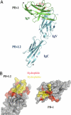
Fig. 1.
Fig. 1.
Structure of the PD-1/PD-L2 complex. (A) Overall structure of the PD-1/PD-L2 complex. Green, PD-1; cyan, PD-L2. The strands of PD-1 and PD-L2 are labeled in red and blue, respectively. (B) Surface representation of PD-1/PD-L2 binding interface. Red, hydrophilic residues in the binding interface; yellow, hydrophobic residues in the binding interface. PD-L2 is in the same orientation as in A; PD-1 is rotated 180° about a vertical axis to reveal the binding surface.
Fig. 2.
Fig. 2.
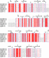
Alignment of the PD-1 ectodomains. The β strands in mouse PD-1 are denoted with arrow segments above the sequence. Red shading, conserved residues; red labeling, residues with similar properties; green triangles, residues bearing potential N-glycans; green asterisks, residues that contribute to binding to PD-L2.
Fig. 3.
Fig. 3.
Alignment of the extracellular domains of PD-L1 and PD-L2. The β strands in mouse PD-L2 are denoted with arrows above the sequence. Red shading, conserved residues; red labeling, residues with similar properties; triangles, residues bearing potential N-glycans; pink, residues conserved between PD-Ls; green, residues conserved for PD-L2 only; green asterisks, residues that contribute to receptor binding of PD-L2; filled circles, residues forming the interdomain hydrogen bonds between PD-L2 IgV and IgC domains.
Fig. 4.
Fig. 4.
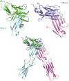
Comparison of the PD-1/PD-L2 and the CTLA-4/B7–1 complexes. (Upper Left) IgV domains of PD-1 (green) and PD-L2 (cyan) in the PD-1/PD-L2 complex. (Upper Right) IgV domains of CTLA-4 (blue) and B7–1 (magenta) in the CTLA-4/B7–1 complex. (Lower) Overlay of the entire PD-1/PD-L2 and CTLA-4/B7–1 complexes by superimposition of PD-1 and CTLA-4.
Fig. 5.
Fig. 5.
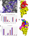
Mapping the binding interface between PD-1 and PD-L2. (A) The core of the binding interface: electron density of residues W110 through Y114 from the G strand of PD-L2 contacting the front concave surface of PD-1 formed by the C, F, and G strands. (B) Receptor binding interface of PD-L2. Mutation of those residues shown in red results in significantly reduced or no binding. Other residues predicted to form the PD-L2 binding site based on the structure of the complex are shown in magenta. Mutation of those residues shown in gray had little or no effect on binding to PD-1. (C) Binding of cell surface expressed PD-L2 mutants (DW110 indicates deletion of residue W110) to PD-1 Ig (0.5 and 5 μg/ml), detected by flow cytometry. Data are shown as percentages of MFI values of wild-type PD-L2 binding to PD-1. (D) Ligand binding surface of PD-1. Mutation of those residues shown in red results in significantly reduced or no binding to either of the ligands. Mutation of those residues shown in blue decreases binding to PD-L1 only, but not to PD-L2. Mutation of A99 (green) increases affinity for both ligands. Other residues contributing to the ligand-binding site based on the structure of the complex are shown in magenta. Mutation of those residues shown in gray had little or no significant effect on binding to PD-L2. (E) Binding of PD-1 mutants to PD-L1 (blue) and PD-L2 (purple) Ig. Data are shown as percentages of MFI values of wild-type PD-1 binding to either PD-L1 or PDL-L2.
Fig. 6.
Fig. 6.
PD-1/PD-L interaction at the cell–cell interface. Noncovalent interactions between PD-1 and PD-Ls are sufficient to drive their enrichment at a pseudosynapse. (A and B) PD-1 and PD-L1 (A) or PD-L2 (B) expressed in CHO cells are recruited to the cell–cell contact area and form conjugates that are analogous to the immunological synapse. (Left) PD-1-CFP-expressing cells in blue. (Center) PD-L1-YFP or PD-L2-YFP-expressing cells in yellow. (Right) Overlay of the CFP and YFP images. (C) Model of the PD-1/PD-L2 complex in the immunological synapse. A number of receptor–ligand assemblies have dimensions that are compatible with colocalization to the central zone of the immunological synapse.

VSports app下载 - Comment in

References

    1. Greenwald RJ, Freeman GJ, Sharpe AH. The B7 family revisited. Annu Rev Immunol. 2005;23:515–548. - PubMed
    1. Bhatia S, Edidin M, Almo SC, Nathenson SG. Different cell surface oligomeric states of B7–1 and B7–2: Implications for signaling. Proc Natl Acad Sci USA. 2005;102:15569–15574. - PMC - PubMed
    1. Okazaki T, Honjo T. PD-1 and PD-1 ligands: From discovery to clinical application. Int Immunol. 2007;19:813–824. - V体育2025版 - PubMed
    1. Freeman GJ, et al. Engagement of the PD-1 immunoinhibitory receptor by a novel B7 family member leads to negative regulation of lymphocyte activation. J Exp Med. 2000;192:1027–1034. - PMC - PubMed
    1. Latchman Y, et al. PD-L2 is a second ligand for PD-1 and inhibits T cell activation. Nat Immunol. 2001;2:261–268. - PubMed

Publication types (V体育ios版)

"VSports app下载" MeSH terms