Skip to main page content
U.S. flag

An official website of the United States government

Dot gov

The VSports app下载. gov means it’s official. Federal government websites often end in . gov or . mil. Before sharing sensitive information, make sure you’re on a federal government site. .

Https

The site is secure V体育官网. The https:// ensures that you are connecting to the official website and that any information you provide is encrypted and transmitted securely. .

. 2008 Sep 5;283(36):25046-56.
doi: 10.1074/jbc.M803776200. Epub 2008 Jul 3.

"V体育官网" Human AlkB homolog 1 is a mitochondrial protein that demethylates 3-methylcytosine in DNA and RNA

Affiliations

Human AlkB homolog 1 is a mitochondrial protein that demethylates 3-methylcytosine in DNA and RNA

Marianne Pedersen Westbye et al. J Biol Chem. .

V体育官网入口 - Abstract

The Escherichia coli AlkB protein and human homologs hABH2 and hABH3 are 2-oxoglutarate (2OG)/Fe(II)-dependent DNA/RNA demethylases that repair 1-methyladenine and 3-methylcytosine residues. Surprisingly, hABH1, which displays the strongest homology to AlkB, failed to show repair activity in two independent studies. Here, we show that hABH1 is a mitochondrial protein, as demonstrated using fluorescent fusion protein expression, immunocytochemistry, and Western blot analysis. A fraction is apparently nuclear and this fraction increases strongly if the fluorescent tag is placed at the N-terminal end of the protein, thus interfering with mitochondrial targeting. Molecular modeling of hABH1 based upon the sequence and known structures of AlkB and hABH3 suggested an active site almost identical to these enzymes. hABH1 decarboxylates 2OG in the absence of a prime substrate, and the activity is stimulated by methylated nucleotides. Employing three different methods we demonstrate that hABH1 demethylates 3-methylcytosine in single-stranded DNA and RNA in vitro. Site-specific mutagenesis confirmed that the putative Fe(II) and 2OG binding residues are essential for activity VSports手机版. In conclusion, hABH1 is a functional mitochondrial AlkB homolog that repairs 3-methylcytosine in single-stranded DNA and RNA. .


Figures

FIGURE 1.
FIGURE 1.
Oxidative demethylation of 3-meC in DNA/RNA catalyzed by AlkB. The co-substrate 2OG is decarboxylated, whereas the methyl group is oxidized to hydroxymethyl and subsequently released as formaldehyde.
FIGURE 2.
FIGURE 2.
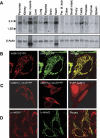
Tissue distribution and subcellular localization of hABH1. A, multiple tissue Northern blots (Clontech, numbers 7759-1/7760-1) were hybridized with 32P-end-labeled full-length cDNA probes for hABH1 and β-actin. In heart and skeletal muscle there are two transcripts of β-actin. B, hABH1-EYFP co-expressed with mitochondrial UNG1-ECFP in HeLa cells. C, subcellular distribution patterns of hABH1-(1-119)-EYFP, hABH1-(27-389)-EYFP, and EYFP-hABH1. D, immunostaining of HeLa cells with monoclonal anti-hABH1 and polyclonal anti-mitochondrial antiviral signaling protein (MAVS) antibodies.
FIGURE 3.
FIGURE 3.
Western blot analyses of subcellular fractions. The same amount of protein extracts (typically 20 μg) from different fractions as indicated were subjected to Western blot analysis using monoclonal antibodies against hABH1, hABH2, and PCNA, and polyclonal antibodies against VDAC (outer mitochondrial membrane marker) and COX (mitochondrial matrix marker) as indicated. A, subcellular fractionation and identification of hABH1 and hABH2 in mitochondria and nuclei, respectively. B, freshly prepared mitochondrial fractions were treated with various amounts of Proteinase K, centrifuged to remove degraded proteins, and the pellet was subjected to Western blot analysis. As a control recombinant hABH1(Δ3) was subjected to the same treatment and analyzed by SDS-PAGE. C, the mitochondria were treated with digitonin, and centrifuged to separate extracted components. The same amounts of supernatant (S) and pellet (P) were analyzed by Western blotting.
FIGURE 4.
FIGURE 4.
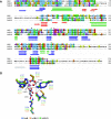
Structure sequence-based alignment of AlkB, hABH1, and hABH3 and three-dimensional model prediction for hABH1 active site residues. A, sequence-structure alignment of AlkB, hABH1, and hABH3 generated using ClustalX (43) based on crystal structures of AlkB (2FD8) and hABH3 (2IUW) and secondary structure of hABH1, predicted by the program JNet (44). The alignment was edited with JalView (45) and the sequence colored according to the ClustalX color scheme showing the degree of conservation. The active site residues subjected to analyses in the present study comprise the iron-binding motif (asterisks), 2OG-binding motif (circles), as well as Ile218 in hABH1 (arrowhead). α-Helices (cylinders) and β-strands (arrows) are colored green and blue, respectively, for conserved regions, and red for the variable subdomain forming part of the DNA/RNA-binding groove. A putative α-helix only present in hABH1 is colored gray. B, close-up view of the predicted active site of hABH1 (orange) superimposed onto hABH3 (2IUW) (green) and AlkB (2FD8) (blue). A three-dimensional model of the central catalytic core domain of hABH1 was predicted by the automated comparative homology modeling server SWISS_MODEL (46) based on the structure-sequence alignment presented in Fig. 4A using the crystal structure of AlkB as template. The side chains of the iron- and 2OG-binding motifs of the respective protein, as well as 2OG (hABH3), are represented as balls and sticks. The partly oxidized Leu177 in hABH3 is denoted with an asterisk and the polar contacts between active site atoms as dotted lines. The iron ion (magenta) and the iron-bound water (red) are shown as spheres.
FIGURE 5.
FIGURE 5.
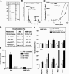
2OG decarboxylase activity of hABH1(Δ3). A, SDS-PAGE of recombinant hABH1(Δ3) and His-tagged AlkB purified to apparent homogeneity. B, mass shift of 16 Da for the peptide 214AEAGILNYYR223 of hABH1. C, AlkB or hABH1(Δ3) were incubated with 1-[14C]2OG (∼24,000 dpm). The released 14CO2 was collected and analyzed by scintillation counting. D, AlkB (10 pmol) and hABH1(Δ3) (30 pmol) were assayed for release of 14CO2 from 1-[14C]2OG when Fe(II) was either absent or chelated by adding 1 mm EDTA. E, effect of the methylated nucleosides 1-methyladenosine (1-meAde), 6-methyladenosine (6-meAde), and 3-methylcytidine (3-meCyt) on 2OG decarboxylation by hABH1(Δ3) and AlkB (15 pmol). F, effect of MMS-treated and untreated DNA and RNA on decarboxylation of 2OG by hABH1(Δ3) and AlkB (15 pmol).
FIGURE 6.
FIGURE 6.
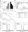
Demethylase activity of hABH1(Δ3). A, demethylase activity of hABH1(Δ3) (10 pmol), hABH3 (10 pmol), and AlkB (5 pmol) on different [3H]methylnitrosourea-methylated polymers of DNA and RNA. B, [3H]methyl-poly(dC) was incubated with AlkB or hABH1(Δ3) or with both proteins for 1 h, followed by ethanol precipitation. The ethanol-soluble radioactivity was quantified by scintillation counting. C, inhibition by polyclonal anti-hABH1 antibody of hABH1(Δ3) (15 pmol) demethylase activity using [3H]methyl-poly(dC) as substrate. AlkB (15 pmol) was used as control. D, demethylase activity of hABH1(Δ3) (20 pmol), on single-stranded oligonucleotide substrate (1 pmol) containing 3-meC, using the fluorescent oligonucleotide substrate assay. The standard reaction mixture was modified as depicted. E, HPLC elution profile of the products generated by acid hydrolysis of the ethanol-precipitated [3H]methyl-poly(dC) after incubation with AlkB or hABH1(Δ3) (10 pmol), or without enzyme. The arrow indicates retention time for unlabeled 3-meC standard as detected by UV absorbance at 260 nm. F, as in E but including whole mitochondria cell extract (MPE) (50 μg), hABH1(Δ3) (10 pmol), or without enzyme.
FIGURE 7.
FIGURE 7.
Repair of 1-meA and 3-meC by hABH1(Δ3). A, DNA oligonucleotides containing 1-me(dA) or 3-me(dC) were incubated with hABH3 or hABH1(Δ3) (20 pmol) for 15 min, followed by decomposition to nucleosides and LC/MS/MS quantization. The values are means of triplicates. B, methyl-poly(rA) or methylpoly(rC) were incubated with AlkB or hABH1(Δ3) and treated as in A. The values are means of duplicates. C, repair of carboxyfluorescein end-labeled single-stranded or double-stranded 49-mer DNA oligonucleotides containing 3-me(dC) or a 1-me(dA). The substrates were incubated with hABH1(Δ3) (0-20 pmol) or AlkB (10 pmol) and subsequently cleaved with DpnII restriction enzyme, run on PAGE, and analyzed for fluorescence.
FIGURE 8.
FIGURE 8.
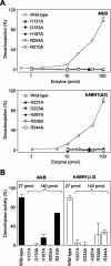
Analysis of active site residues in AlkB and hABH1(Δ3) involved in iron and 2OG binding. A, 2OG decarboxylase activity of AlkB, hABH1(Δ3), and mutants in the iron-binding motif (H131A/H233A, D131A/D233A, H187A/H287A) and 2OG-binding motif (R204A/2338A, R210A/R344A). B, demethylase activity of wild-type and various mutants of AlkB and hABH1(Δ3) on [3H]methyl-poly(dC) substrate. Note that the mutant proteins were added in excess (∼5-fold) as compared with the wild-type proteins.

"VSports手机版" References

    1. Drabløs, F., Feyzi, E., Aas, P. A., Vaagbø, C. B., Kavli, B., Bratlie, M. S., Peña-Diaz, J., Otterlei, M., Slupphaug, G., and Krokan, H. E. (2004) DNA Repair 31389 -1407 - PubMed
    1. Sedgwick, B. (2004) Nat. Rev. Mol. Cell Biol. 5148 -157 - PubMed (VSports注册入口)
    1. Sedgwick, B., and Lindahl, T. (2002) Oncogene 218886 -8894 - PubMed
    1. Kataoka, H., Yamamoto, Y., and Sekiguchi, M. (1983) J. Bacteriol. 1531301 -1307 - PMC - PubMed
    1. Falnes, P. O., Johansen, R. F., and Seeberg, E. (2002) Nature 419178 -182 - VSports app下载 - PubMed

VSports手机版 - Publication types

MeSH terms

VSports在线直播 - Substances