Skip to main page content
U.S. flag

An official website of the United States government

Dot gov

The VSports app下载. gov means it’s official. Federal government websites often end in . gov or . mil. Before sharing sensitive information, make sure you’re on a federal government site. .

Https

The site is secure V体育官网. The https:// ensures that you are connecting to the official website and that any information you provide is encrypted and transmitted securely. .

. 2008 Jul 1;22(13):1838-50.
doi: 10.1101/gad.466308.

Translational regulation of glutathione peroxidase 4 expression through guanine-rich sequence-binding factor 1 is essential for embryonic brain development

Affiliations

Translational regulation of glutathione peroxidase 4 expression through guanine-rich sequence-binding factor 1 is essential for embryonic brain development

Christoph Ufer et al. Genes Dev. .

Abstract

Phospholipid hydroperoxide glutathione peroxidase (GPx4) is a moonlighting selenoprotein, which has been implicated in basic cell functions such as anti-oxidative defense, apoptosis, and gene expression regulation. GPx4-null mice die in utero at midgestation, and developmental retardation of the brain appears to play a major role. We investigated post-transcriptional mechanisms of GPx4 expression regulation and found that the guanine-rich sequence-binding factor 1 (Grsf1) up-regulates GPx4 expression. Grsf1 binds to a defined target sequence in the 5'-untranslated region (UTR) of the mitochondrial GPx4 (m-GPx4) mRNA, up-regulates UTR-dependent reporter gene expression, recruits m-GPx4 mRNA to translationally active polysome fractions, and coimmunoprecipitates with GPx4 mRNA. During embryonic brain development, Grsf1 and m-GPx4 are coexpressed, and functional knockdown (siRNA) of Grsf1 prevents embryonic GPx4 expression. When compared with mock controls, Grsf1 knockdown embryos showed significant signs of developmental retardations that are paralleled by apoptotic alterations (TUNEL staining) and massive lipid peroxidation (isoprostane formation). Overexpression of m-GPx4 prevented the apoptotic alterations in Grsf1-deficient embryos and rescued them from developmental retardation. These data indicate that Grsf1 up-regulates translation of GPx4 mRNA and implicate the two proteins in embryonic brain development VSports手机版. .


Figures
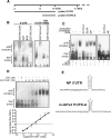
Figure 1.
Figure 1.
Grsf1 specifically binds to the 5′UTR of the m-GPX4 mRNA. Protein binding to the 5′UTR of m-GPx4 mRNA was studied by RNA mobility gel shift assays. For this purpose, two labeled RNA probes representing different parts of the 5′UTR of m-GPx4 mRNA were incubated in vitro with different amounts of purified recombinant K6-Grsf1/GST fusion proteins. Aliquots of this incubation mixture were loaded on a 5% polyacrylamide gel (native conditions) and the separated protein–RNA complexes were then transferred to a nylon membrane. The blots were visualized as described in the Materials and Methods. (A) 5′UTR of the m-GPx4 mRNA and RNA probes (5′UTR and 5′UTR-A) used for initial shift assays. The 5′-ATG represents the start codon for m-GPx4 and the 3′-ATG that for the cytosolic enzyme. The sequence between these two ATG codons represents the mitochondrial insertion sequence. Letters a and b indicate the position of the two major transcription initiation sites for m-GPx4 (Nam et al. 1997; Knopp et al. 1999). (B) Biotin-labeled probes of the m-GPx4 5′UTR or 18S rRNA (negative control) were incubated with 4.5 μg of the indicated proteins. (C) Digoxigenin-labeled probes of m-GPx4 5′UTR (5′UTR-A), influenza NP 5′UTR (NP), or SEAP 5′UTR (SEAP) were incubated with K6-Grsf1/GST fusion protein in the absence or presence of 50 pmol of unlabeled competitor RNA. (D, top part) A digoxigenin-labeled probe, 5′UTR-A, was incubated with varying amounts of free GST or Grsf1/GST fusion protein. (Bottom part) The ratio of signal intensities of the RNA shift band/free RNA were plotted against Grsf1/GST concentration. The intercept with the X-axis represents the Kd. (E) Sequence and predicted secondary structure of influenza NP 5′UTR (Park et al. 1999) and m-GPx4 5′UTR.
Figure 2.
Figure 2.
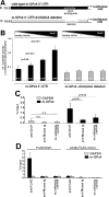
Grsf1 expression up-regulates expression of UTR-dependent reporter gene constructs and interacts with m-GPx4 mRNA in vivo. UTR-dependent reporter gene assays were carried out as described in the Supplemental Material. (A) For co-transfection of embryonic fibroblasts with Grsf1, two different luciferase-based reporter gene constructs containing the 5′UTR of m-GPx4 were designed. One construct contained the consensus Grsf1-binding sequence (wild-type m-GPx4 5′UTR), and the other lacked this motif (m-GPx4 5′UTR AGGGGA deletion). Increasing concentrations of Grsf1 expression plasmid were co-transfected with the reporter gene constructs. To adjust a constant quantity of pGL3-promoter, all samples were supplemented with empty vector (the proportion of Grsf1 construct was 0%, 25%, 50%, and 100%, respectively, as indicated by the bar graphics above the diagram). After co-transfection, cells were kept in culture for 6 h and luciferase activity was measured in the lysates. (B) The relative luciferase activities (numbers at the bottom of each column) were calculated. The activities measured were corrected for transfection efficiency (Renilla luciferase activity) and normalized to equal amounts of pGL3-promoter. (C) RNA immunoprecipitation in murine N2a cells was carried out as described in the Supplemental Material. RNA recovered from “Input” samples was set at 100%. Data are given as means of three independent experiments ±SD (Student’s t-test). (D) RNA immunoprecipitations in N2a cells transfected with a Grsf1/Flag expression plasmid or an empty Flag vector were carried out as described in the Supplemental Material. RNA recovered from “Input” samples was set at 100%. Data are given as means of three independent experiments ±SD.
Figure 3.
Figure 3.
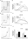
Polysomal gradient analysis. MEF cells were transiently transfected for 24 h using either a Grsf1 expression or a control vector (no Grsf1 insert). (A–C) Typical polysome profiles after sucrose gradient ultracentrifugation monitored at 254 nm from the bottom (51% sucrose, left) to top (17% sucrose, right) are shown. Each gradient was separated into 12 fractions. The ribosomal profile in C was determined in the presence of 25 mM EDTA. (D) Quantification of m-GPx4 mRNA concentration per fraction by RT–PCR. (E) β-Actin mRNA levels per fraction according to D. (F) Statistical evaluation of m-GPx4 mRNA levels summarized for polysomal and nonpolysomal fractions. Grsf1 overexpression resulted in a shift of m-GPx4 mRNA into polysomal fractions. Mean values are given and error bars represent the standard deviation. n = 3; (*) P < 0.05.
Figure 4.
Figure 4.
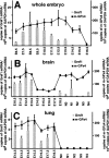
Grsf1 and m-GPx4 are coexpressed during murine embryogenesis. Murine embryos were prepared from pregnant mice at different developmental stages E6.5–E18.5 and postnatal stages N0–N4, and total RNA was extracted. Steady-state concentrations of Grsf1 and m-GPx4 mRNA were quantified by qRT–PCR using GAPDH as an internal standard. To explore expression of the two genes during brain and lung development, these organs were prepared at different developmental stages and the two mRNA species were quantified in total RNA extracts. The numbers indicate the days of gestation. E6.5 means 6.5 d post-conception. N0 indicates the day of birth, and N1 means 24 h after birth.
Figure 5.
Figure 5.
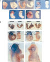
Targeted knockdown of Grsf1 impairs expression of m-GPx4 during murine embryogenesis and induces developmental retardation. Mouse embryos were prepared at gestational day E8, treated with control siRNA duplex (labeled as Control) or Grsf1-specific siRNA constructs (labeled as siRNA), and then cultured in vitro for up to 72 h. After different time points of the culturing period, the embryos were used for in situ hybridization using Grsf1- and m-GPx4-specific antisense probes. Dark areas indicate regions with intense hybridization signals. The different panels represent different stages of embryonic development at the end of the in vitro culturing period. Each panel consists of a left image (control embryo) and a right image (siRNA-treated embryo). Bar, 300 μm (E8–E8.5); 800 μm (E9.5–E10). (hf) Headfold; (tb) tailbud; (fb) forebrain; (mb) midbrain; (hb) hindbrain; (r) rhombomere. (Panel I) Grsf1 in situ hybridization. siRNA treatment induced abnormal mid/hindbrain development (red arrows indicate retarded mid/hindbrain boundary), posterior truncation (red dotted areas indicate shorted and twisted tail bud), and general growth retardation (white arrows indicate shorted crl). (Panel II) m-GPx4 in situ hybridization. siRNA treatment impaired m-GPx4 mRNA expression from E9.5 and induced retarded hindbrain segments at r5/6 levels.
Figure 6.
Figure 6.
m-GPx4 overexpression rescues developmental abnormalities induced by Grsf1 knockdown. Grsf1 siRNA-treated embryos (E7.5) were transfected with a mammalian overexpression vector containing or lacking (control) the m-GPx4 coding sequence and then cultured in vitro for 48 h. Then the embryos were recovered and analyzed. (Panel I) Quantification of developmental characteristics according to the scoring procedure described in the Supplemental Material. (Panel II) In situ hybridization of cotransfected embryos using a m-GPx4 specific probe. Each panel consists of a left image (control embryo) and a right image (siRNA treated embryo). (Panel III) The embryos were sectioned for immunohistochemistry analysis using a specific m-GPx4 antibody. Positive staining is indicated as dark brown areas. Each panel consists of left images (no vector) and right images (m-GPx4 overexpression vector). (Right panels) Magnified neuroepithelium regions from the brain are shown. Shown are mean values and error bars represent the standard deviation. n = 6; (*) P < 0.001.
Figure 7.
Figure 7.
m-GPx4 overexpression reduces cerebral lipid peroxidation and apoptosis induced by Grsf1 knockdown. Grsf1 siRNA-treated embryos (E7.5) were transfected with a mammalian overexpression vector containing or lacking (control) the m-GPx4 coding sequence. After a culturing period of 48 h, the embryos were recovered and analyzed for lipid peroxidation (isoprostane content, A) and apoptosis (TUNEL assay, B). Shown are mean values, and error bars represent the standard deviation. n = 5; (*) P < 0.01.

References

    1. Altschul S.F., Madden T.L., Schaffer A.A., Zhang J., Zhang Z., Miller W., Lipman D.J. Gapped BLAST and PSI-BLAST: A new generation of protein database search programs. Nucleic Acids Res. 1997;25:3389–3402. - PMC - PubMed
    1. Bernstein D.S., Buter N., Stumpf C., Wickens M. Analyzing mRNA–protein complexes using a yeast three-hybrid system. Methods. 2002;26:123–141. - PubMed
    1. Birney E., Kumar S., Krainer A.R. Analysis of the RNA-recognition motif and RS and RGG domains: Conservation in metazoan pre-mRNA splicing factors. Nucleic Acids Res. 1993;21:5803–5816. - PMC - PubMed
    1. Borchert A., Savaskan N.E., Kuhn H. Regulation of expression of the phospholipid hydroperoxide/sperm nucleus glutathione peroxidase gene. Tissue-specific expression pattern and identification of functional cis- and trans-regulatory elements. J. Biol. Chem. 2003;278:2571–2580. - PubMed
    1. Borchert A., Wang C.C., Ufer C., Schiebel H., Savaskan N.E., Kuhn H. The role of phospholipid hydroperoxide glutathione peroxidase isoforms in murine embryogenesis. J. Biol. Chem. 2006;281:19655–19664. - PubMed

V体育安卓版 - Publication types

V体育ios版 - MeSH terms

V体育2025版 - Substances

V体育安卓版 - LinkOut - more resources