Skip to main page content
U.S. flag

An official website of the United States government

Dot gov

The . gov means it’s official. Federal government websites often end in . gov or VSports app下载. mil. Before sharing sensitive information, make sure you’re on a federal government site. .

Https

The site is secure V体育官网. The https:// ensures that you are connecting to the official website and that any information you provide is encrypted and transmitted securely. .

. 2008 Aug 15;112(4):1424-33.
doi: 10.1182/blood-2008-01-133769. Epub 2008 Jun 9.

Vorinostat inhibits STAT6-mediated TH2 cytokine and TARC production and induces cell death in Hodgkin lymphoma cell lines

Affiliations

Vorinostat inhibits STAT6-mediated TH2 cytokine and TARC production and induces cell death in Hodgkin lymphoma cell lines

VSports注册入口 - Daniela Buglio et al. Blood. .

Abstract

Epigenetic changes have been implicated in silencing several B-cell genes in Hodgkin and Reed-Sternberg cells (HRS) of Hodgkin lymphoma (HL), and this mechanism has been proposed to promote HRS survival and escape from immunosurveillance. However, the molecular and functional consequences of histone deacetylase (HDAC) inhibition in HL have not been previously described VSports手机版. In this study, we report that the HDAC inhibitor vorinostat induced p21 expression and decreased Bcl-xL levels causing cell-cycle arrest and apoptosis. Furthermore, vorinostat inhibited STAT6 phosphorylation and decreased its mRNA levels in a dose- and time-dependent manner, which was associated with a decrease in the expression and secretion of Thymus and Activation-Regulated Chemokine (TARC/CCL17) and interleukin (IL)-5 and an increase in IP-10 levels. Moreover, vorino-stat inhibited TARC secretion by dendritic cells that were activated by the thymic stromal lymphopoietin (TSLP). Collectively, these data suggest that pharmacologic HDAC inhibition in HL may induce favorable antitumor activity by a direct antiproliferative effect on HRS cells, and possibly by an immune mediated effect by altering cytokine and chemokines secretion in the microenvironment. .


Figures
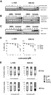
Figure 1
Figure 1
Effect of HDAC inhibitor vorinostat in Hodgkin lymphoma (HL) cell lines. (A) Cells were incubated with 0.1 to 5 μM of vorinostat for 6 hours and whole cell lysates were examined for histone 3 acetylation (Ac-histone 3) using Western blot analysis. During this time frame, histone acetylation was observed using concentrations of 2 μM or higher. (B) Cells were incubated with 5 μM of vorinostat for 0.5 to 6 hours demonstrated that histone acetylation is achieved as early as 0.5 hours of incubation and lasted for at least 6 hours. (C) Vorinostat (5 μM) induced p21 expression as early as 6 hours and lasted for up to 48 hours. (D) Vorinostat induced antiproliferative effects in HL cell lines as determined by the MTS cell proliferation assay. This effect was prominent after 48 hours in culture and was dose dependent. Each value is the mean of 3 independent experiments performed in triplicate (± SEM). (E) The antiproliferative effect of vorinostat was associated with an increase in the G2/M cell-cycle fraction, especially when higher concentrations were used. The G2M fraction was higher after 48 hours of incubation (bottom panel) compared with 24 hours of incubation (top panel). Results are the means of 3 independent experiments.
Figure 2
Figure 2
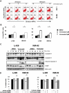
Vorinostat induces apoptosis in HL cell lines. (A) A representative experiment demonstrating the effect of 2 different concentrations of vorinostat (1 and 5 μM) on apoptosis as determined by the annexin-V binding assay. HL cell lines were incubated with medium or vorinostat (1-5 μM) for 24 or 48 hours before the percentage of dead cells was determined using dual staining with annexin-V and propidium iodide. Vorinostat induces apoptosis in both HL cell lines especially when higher concentrations were used for 48 hours. Percentages of dead cells are shown in each quadrant. (B) Summary results of vorinostst-induced cell death (PI and annexin-V positive cells) from 3 independent experiments. Results after incubations for 24 hours (left panel) and 48 hours (right panel) are shown. Each value is the mean of 3 independent experiments performed in triplicate (± SEM). * denotes a P value of less than .05, and ** denotes a P value of less than .005. (C) HL cell lines were incubated with medium or 5 μM of vorinostat for 6 to 48 hours. Whole cell lysates were examined by Western blot for changes in intracellular proteins. Using this high concentration of vorinostat (5 μM) apoptosis was associated with activation of caspases 8, 9, 3 and PARP cleavage. Consistent with data presented in Figure 2A and B, lower concentrations of vorinostat (1 μM) did not induce caspase activation during the same time frame (Figure S1, available on the Blood website; see the Supplemental Materials link at the top of the online article). (D) Vorinostat-induced cell death of HL cell lines was either partially or completely blocked by the pan-caspase inhibitor Z-VAD-FMK (left panel) or by the caspase 9 inhibitor Z-LEHD-FMK (right panel). HL cell lines were incubated with medium or Z-VAD-FMK (20 μM), Z-LEHD-FMK (20 μM), vorinostat (5 μM), or a combination of vorinostat and Z-VAD-FMK or vorinostat and Z-LEHD-FMK. After 24 hours, the viable cells were counted using a 3-(4,5dimethyltioazol-2-yl)-5-(3-carboxymethoxy-phenyl)-2-(4-sulfonyl)-2H-tetrazolium (MTS) assay. Each value represents a mean of 3 independent experiments done in triplicate (± SEM). * denotes a P value of less than .05.
Figure 3
Figure 3
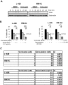
Effect of vorinostat inhibits STAT6 in HL cell lines. (A) L-428 and KM-H2 cell lines were incubated with 5 μM of vorinostat for 6 to 48 hours. Whole cell lysates were examined by Western blotting. As shown, vorinostat induced down-regulation of pAkt (Ser473) without inducing changes in total Akt or pAkt (Thr308), and decreased STAT6 and pSTAT6 levels without a significant effect on STAT3. (B) Vorinostat down-regulated STAT6 mRNA expression in a dose-dependent manner. Cells were cultured with medium and vorinostat (1-5 μM) for 24 hours, and the effects on STAT6 mRNA expression was determined by the TaqMan PCR assay. Results were normalized to GAPDH and represent the mean of 3 independent experiments (± SEM). (C) L428 cell line was transfected with STAT6 siRNA and the efficacy of this method was confirmed by measuring STAT6 protein levels by Western blot. Cells that were incubated with STAT6 siRNA demonstrated a 50% reduction in STAT6 protein after 48 hours compared with those incubated with control siRNA. This is a representative experiment of 3 independent experiments demonstrating similar results. (D) Effect of STAT6 knock down by siRNA on cell proliferation. L-428 cells that were transfected with STAT6 siRNA demonstrated lower proliferative rate and thymidine incorporation compared with those transfected with control siRNA.
Figure 4
Figure 4
Effect of vorinostat on TH1/TH2 cytokine secretion in HL cell lines. Cells were incubated with medium or vorinostat (5 μM) for 24 hours and supernatants were examined for cytokine levels by ELISA. Each value represents a mean of 3 independent experiments done in triplicate (± SEM).
Figure 5
Figure 5
Regulation of TARC by vorinostat. (A) Vorinostat inhibits TARC mRNA expression in HL cell lines. Cells were incubated with increasing concentrations of vorinostat for 24 hours before TARC mRNA levels were determined by RT-PCR as detailed in Methods section. Each value represents a mean of 3 independent experiments done in triplicate (± SEM). (B) Vorinostat decreased TARC secretion in L-428 and KM-H2. Cells were incubated with vorinostat 2 μM for 24 to 48 hours before TARC levels were determined in the supernatants by ELISA. Each value represents a mean of 3 independent experiments done in triplicate (± SEM). (C) Silencing STAT6 by siRNA decreases TARC secretion in the L-428 cells. Cells were transfected with STAT6 siRNA or control siRNA for 12 to 72 hours. At each time point, culture supernatants were collected and TARC levels were determined by ELISA. Each value represents a mean of 3 independent experiments done in triplicate (± SEM). (D) Vorinostat inhibits TARC secretion from DCs collected from 2 healthy donors and activated with TSLP (100 ng/mL). TARC levels were determined in DC supernatants after 24 hours in culture by ELISA. The right and left panels represent 2 independent experiments using DCs from 2 healthy donors.
Figure 6
Figure 6
Vorinostat decresases Bcl-xL protein levelsand enhances the effect of doxorubicin and gemcitabine chemotherapy in HL. (A) Vorinostat (5 μM) decreases the level of the antiapoptotic protein Bcl-xL in HL cell lines in a time dependent manner as determined by Western blot. (B) Representative experiments demonstrating synergistic effects between vorinostat and doxorubicin (left panel) and gemcitabine (right panel). Cells were incubated with each drug alone or in combination for 48 hours and cell viability was determined by the MTS assay. Synergy was determined by calculating the combination index (CI) analyzed by Calcusyn software. Each value represents a mean of 3 independent experiments performed in triplicate (± SEM). (C) Synergy between Vorinostat and gemcitabine (top table) or doxorubicin (bottom table) using a range of drug concentrations. Synergy was determined by calculating the CI using the Calcusyn software. A CI less than 1 indicates synergy.
Figure 7
Figure 7
A model for vorinostat activity in HL. HL is characterized by the presence of abundant numbers of dendritic cells (DCs) and Th2-type CD4 cells in the microenvironment. HRS cells aberrantly express activated STAT6 by an autocrine IL-13 loop. Both DC and HRS cells secrete TARC that attracts TH2 type cells. In this model, vorinostat has a dual effect on HL. (A) A direct effect on HRS cells by modulating a variety of growth and survival proteins, including p21 and Akt, in addition to inhibiting STAT6 transcription. Inhibition of STAT6 decreases Bcl-xL protein levels causing apoptosis, and decreases TARC secretion causing inhibition of Th2-type CD4+ cell chemotaxis. (B) An indirect effect on DCs in the microenvironment. Thymic stromal lymphopoietin (TSLP) activates myeloid DCs (mDCs) to express OX40 ligand (OX40L) and to secrete TARC, favoring polarization and attraction of TH2-type CD4+ cells. Vorinostat inhibits TRAC secretion by DCs, thus modulating the function and component of the reactive cells in HL microenvironment.

VSports app下载 - References

    1. Skinnider BF, Mak TW. The role of cytokines in classical Hodgkin lymphoma. Blood. 2002;99:4283–4297. - V体育安卓版 - PubMed
    1. Re D, Thomas RK, Behringer K, Diehl V. From Hodgkin disease to Hodgkin lymphoma: biologic insights and therapeutic potential. Blood. 2005;105:4553–4560. - PubMed (VSports注册入口)
    1. Baus D, Pfitzner E. Specific function of STAT3, SOCS1, and SOCS3 in the regulation of proliferation and survival of classical Hodgkin lymphoma cells. Int J Cancer. 2006;118:1404–1413. - PubMed
    1. Cochet O, Frelin C, Peyron JF, Imbert V. Constitutive activation of STAT proteins in the HDLM-2 and L540 Hodgkin lymphoma-derived cell lines supports cell survival. Cell Signal. 2006;18:449–455. - PubMed
    1. Skinnider BF, Elia AJ, Gascoyne RD, et al. Signal transducer and activator of transcription 6 is frequently activated in Hodgkin and Reed-Sternberg cells of Hodgkin lymphoma. Blood. 2002;99:618–626. - PubMed

Publication types

MeSH terms