Skip to main page content
U.S. flag

An official website of the United States government

Dot gov

The . gov means it’s official. Federal government websites often end in . gov or VSports app下载. mil. Before sharing sensitive information, make sure you’re on a federal government site. .

Https

The site is secure. The https:// ensures that you are connecting to the official website and that any information you provide is encrypted and transmitted securely. V体育官网.

. 2008 Jun 1;22(11):1522-33.
doi: 10.1101/gad.1652308.

Identification of novel genes involved in light-dependent CRY degradation through a genome-wide RNAi screen

Affiliations

Identification of novel genes involved in light-dependent CRY degradation through a genome-wide RNAi screen

V体育安卓版 - Sriram Sathyanarayanan et al. Genes Dev. .

Abstract

Circadian clocks regulate many different physiological processes and synchronize these to environmental light:dark cycles. In Drosophila, light is transmitted to the clock by a circadian blue light photoreceptor CRYPTOCHROME (CRY). In response to light, CRY promotes the degradation of the circadian clock protein TIMELESS (TIM) and then is itself degraded. To identify novel genes involved in circadian entrainment, we performed an unbiased genome-wide screen in Drosophila cells using a sensitive and quantitative assay that measures light-induced degradation of CRY. We systematically knocked down the expression of approximately 21,000 genes and identified those that regulate CRY stability. These genes include ubiquitin ligases, signal transduction molecules, and redox molecules. Many of the genes identified in the screen are specific for CRY degradation and do not affect degradation of the TIM protein in response to light, suggesting that, for the most part, these two pathways are distinct. We further validated the effect of three candidate genes on CRY stability in vivo by assaying flies mutant for each of these genes. This work identifies a novel regulatory network involved in light-dependent CRY degradation and demonstrates the power of a genome-wide RNAi approach for understanding circadian biology VSports手机版. .


Figures

Figure 1.
Figure 1.
Development of a primary screen to identify novel components involved in light-dependent CRY degradation. (A) Light-dependent degradation of a CRY-luc fusion protein: S2R+ cells were transfected with expression constructs for a Cry-luc fusion protein and Actin-Renilla luciferase. Cells were exposed to light in the presence of DMSO (control) or proteasome inhibitor MG132 (100 μM) for the indicated times, and luciferase activity was monitored. Luciferase activity was normalized to that of cells kept in darkness, set as 1. Average values from three samples are plotted, and the error bars depict standard error of the mean (SEM). The results are representative of two independent experiments. Light-dependent degradation of the CRY-luciferase fusion protein was blocked by proteasome inhibitor MG132. (B) A single point mutation in the flavin-binding pocket of CRY, CRYb, renders it insensitive to light. S2R+ cells were transfected with expression constructs for CRY-luc or CRYb-luc fusion protein in 384-well plates and assayed for luciferase activity after light exposure for 6 h. Luciferase activity from three samples is plotted relative to the CRY activity in transfected cells kept in the dark (set to 100%); error bars represent SEM. In response to light exposure, activity of the CRY-luc fusion protein was reduced by 83% (P-value = 2 × 10⁻⁹), while activity of the CRYb-luc fusion protein was unchanged. Similar results were obtained in three experiments with different cell densities. (C) RNAi of 26S proteasome components blocks light-dependent CRY degradation. S2R+ cells were cotransfected with the CRY-luc expression construct and dsRNAs, as indicated, in 384-well plates. Act-Renilla luc was included as a transfection control. Ninety-six hours post-transfection, cells were exposed to light and luciferase activity was measured as indicated above. Relative luciferase activity from three samples is plotted; error bars represent SEM. The value obtained with dsRNA against gfp in the dark was set as 100% and used for normalization. dsRNA against cry, which dramatically reduced luciferase activity (data not shown), was used as a positive control. RNAi of the 26S proteasome core components Prosβ2 and DmS13 (also called as Rpn11) significantly blocked light-dependent CRY-luc degradation. Similar results were obtained in two independent experiments. (D) A pilot screen demonstrates the validity of the CRY-luc assay. The light-dependent CRY-luc degradation assay was performed in S2R+ cells with 380 randomly picked dsRNAs from the genomic collection. dsRNA against the proteasome component Prosβ2 was used as a positive control; thread dsRNA was included as a control to verify the efficiency of RNAi. Normalized luciferase levels were compared between the dark- and light-treated plates. Z-scores, standard deviations from the plate mean (see the Materials and Methods for details), are plotted. While dsRNA against Prosβ2 significantly blocked light-dependent CRY-luc degradation (Z-score = −6.2), most dsRNAs had no effect. Similar results were obtained in two independent experiments.
Figure 2.
Figure 2.
Results of the primary screen to identify genes required for light-dependent CRY degradation. (A) Correlation of the replicate Z-scores for the primary screen plates. Artificially elevated Z-scores >10 were eliminated from A and B. (B) Scatter plot of the average Z-score from replicate experiments. Each plate contained a positive control, Prosβ2 dsRNA (with Z-score near −5), which was eliminated from the plot. (C) Functional classification of the 173 hits that were reconfirmed by the CRY-luc degradation assay. The genes were assigned to different functional classes based on the GO annotation and plotted.
Figure 3.
Figure 3.
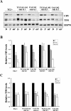
Effect of select candidates on light-dependent CRY and TIM degradation in S2R+ cells. (A) Western blot analysis showing effects of primary screen positives on light-dependent CRY and TIM degradation. S2R+ cells were transfected with expression constructs for cry, tim, and jet and the indicated dsRNA and then either exposed to light for 2 h or maintained in constant darkness (black bar). CRY (top) and TIM (middle) were visualized via Western analysis using V5 and TIM antibodies, respectively. (Bottom) Endogenous HSP-70 levels were used as a loading control. (B) Quantification of the effects of different dsRNAs on CRY degradation. CRY levels were quantified, and differences in average intensities from two experiments were normalized to the dark values and plotted. Error bars represent SEM. Similar results were obtained by using a myc-tagged CRY (data not shown). (C) Quantification of the effects of different dsRNAs on CRY degradation. Relative light-dependent TIM degradation was measured by similar quantification of the TIM band. Based on these values, a relative TIM or CRY degradation score (see the Materials and Methods) was obtained and plotted.
Figure 4.
Figure 4.
Effect of BRUCE on light-dependent CRY and TIM degradation. (A) Bruce mutants have elevated CRY levels that persist even after prolonged light exposure. A representative Western blot image of light-dependent CRY and TIM degradation in yellow white (control) or BruceRB mutants following an acute light pulse (5 min) or prolonged light pulse (60 min) as compared with the dark control (D). High levels of CRY protein were observed in BruceRB mutants, and CRY was not efficiently degraded even after a prolonged light exposure. (B) Western blot analysis of CRY and TIM levels in control and BruceRB mutants in 12:12 light/dark (LD) cycle. Fly heads collected during the light phase (open bar) or dark phase (dark bar) were probed for TIM, CRY, and HSP-70 (loading control). In control flies, CRY levels are high at late night (ZT20), but in BruceRB mutants, CRY levels are low at this time point. Also the overall CRY levels are elevated by more than fivefold in BruceRB mutants. TIM levels are low during the day and high at night in both wild-type and BruceRB mutant flies. (C,D) Quantification of light-dependent CRY and TIM degradation in Bruce mutants. CRY and TIM levels were quantified in the samples described in A and normalized to levels of the respective protein in the control line maintained in the dark. HSP70 levels were determined to control for loading. BruceRB mutants show significantly elevated CRY levels when compared with wild-type siblings and Bruce heterozygotes (P < 0.008). Light-dependent degradation of CRY was also reduced in Bruce mutants (in the data shown here, levels of CRY were significantly reduced in the heterozygous sibling [P < 0.01] but not in the mutant [P = 0.1]). Statistical analysis was conducted using ANOVA followed by a post-hoc Tukey HSD test. An average of four independent experiments is shown.
Figure 5.
Figure 5.
CG17735 regulates light-dependent CRY degradation. (A) Western blot analysis showing the effects of mir 6.1-CG17735 on light-dependent CRY and TIM degradation. Two independent UAS-mir 6.1 CG17735 constructs (541 and 302) were expressed in clock cells using a tim-Gal4 (TG) driver. The TG/UAS-mir 6.1-CG17735, or TG/+ (control) or UAS-mir 6.1-CG17735/CyO (sibling control) flies were treated with either a 5-min light pulse (gray bars) or a 60-min light exposure (white bars) at ZT20 or kept in darkness (black bars). All flies were collected at ZT21. CRY (top) and TIM (middle) were visualized by Western blot analysis using CRY and TIM antibodies, respectively. (Bottom) HSP70 levels were determined to control for loading. Although TIM levels were variable in the 541 line and appear to be higher in the blot shown here, they were generally not affected by knockdown of CG17735. (B,C) Quantification of the effects of CG17735 RNAi on CRY and TIM degradation following a 5- or 60-min light pulse. CRY and TIM levels were quantified, and for each genotype they were normalized to the levels of the respective protein in the dark (D) sample. Error bars represent SEM. Data shown were averaged from four independent experiments for all lines except TG, which is based on three experiments. Post-hoc comparisons (Tukey’s test) show that CRY degradation following the 5-min light pulse is significantly less (P < 0.05) in the CG17735 mir 6.1-expressing lines as compared with the TG control.
Figure 6.
Figure 6.
CRY stability is increased in ssh loss-of-function mutants. (A) Western blot analysis of CRY and TIM degradation in response to an acute light pulse. Flies were given an acute light pulse for 5 min at ZT20, then incubated in the dark and collected at ZT21. (B) Quantification of CRY and TIM levels in the dark-maintained (D) and light-pulsed samples (LP5′). The data represent the average of six biologically independent samples. Levels of CRY were reduced in control samples by 32% (P < 0.01, as compared with the dark control). This light-dependent degradation was significantly less (13%) in ssh mutants (P = 0.09 when compared with its dark control). Although TIM levels were somewhat higher in ssh mutants, its degradation by a light pulse was unaffected (degradation of 47% vs. 42% in control and ssh, respectively). (C) CRY levels are increased in the ssh mutants during the early morning (2 h into the daytime, ZT02). In contrast, TIM levels were low in the daytime and high in the nighttime in both the control and ssh flies. Transheterozygotes of ssh1-6 and ssh51 alleles were used, and Student’s t-test was performed for light-dependent degradation within genotypes.

"VSports app下载" References

    1. Adams J.D., Klaidman L.K., Ribeiro P. Tyrosine hydroxylase: Mechanisms of oxygen radical formation. Redox Rep. 1997;3:273–279. - PubMed (V体育平台登录)
    1. Ashmore L.J., Sehgal A. A fly’s eye view of circadian entrainment. J. Biol. Rhythms. 2003;18:206–216. - PubMed
    1. Bartke T., Pohl C., Pyrowolakis G., Jentsch S. Dual role of BRUCE as an antiapoptotic IAP and a chimeric E2/E3 ubiquitin ligase. Mol. Cell. 2004;14:801–811. - PubMed
    1. Busza A., Emery-Le M., Rosbash M., Emery P. Roles of the two Drosophila CRYPTOCHROME structural domains in circadian photoreception. Science. 2004;304:1503–1506. - PubMed (V体育安卓版)
    1. Cashmore A.R. Cryptochromes: Enabling plants and animals to determine circadian time. Cell. 2003;114:537–543. - "V体育2025版" PubMed

Publication types (V体育安卓版)

"VSports手机版" LinkOut - more resources