Skip to main page content
U.S. flag

An official website of the United States government

Dot gov

The . gov means it’s official. Federal government websites often end in . gov or . mil. Before sharing sensitive information, make sure you’re on a federal government site VSports app下载. .

Https

The site is secure V体育官网. The https:// ensures that you are connecting to the official website and that any information you provide is encrypted and transmitted securely. .

. 2008 Aug;190(15):5480-92.
doi: 10.1128/JB.00384-08. Epub 2008 May 30.

"VSports最新版本" A second pilus type in Streptococcus pneumoniae is prevalent in emerging serotypes and mediates adhesion to host cells

Affiliations

V体育平台登录 - A second pilus type in Streptococcus pneumoniae is prevalent in emerging serotypes and mediates adhesion to host cells

Fabio Bagnoli et al. J Bacteriol. 2008 Aug.

Abstract

Analysis of publicly available genomes of Streptococcus pneumoniae has led to the identification of a new genomic element containing genes typical of gram-positive pilus islets (PIs). Here, we demonstrate that this genomic region, herein referred to as PI-2 (consisting of pitA, sipA, pitB, srtG1, and srtG2) codes for a second functional pilus in pneumococcus. Polymerization of the PI-2 pilus requires the backbone protein PitB as well as the sortase SrtG1 and the signal peptidase-like protein SipA. Presence of PI-2 correlates with the genotype as defined by multilocus sequence typing and clonal complex (CC). The PI-2-positive CCs are associated with serotypes 1, 2, 7F, 19A, and 19F, considered to be emerging serotypes in both industrialized and developing countries VSports手机版. Interestingly, strains belonging to CC271 (where sequence type 271 is the predicted founder of the CC) contain both PI-1 and PI-2, as revealed by genome analyses. In these strains both pili are surface exposed and independently assembled. Furthermore, in vitro experiments provide evidence that the pilus encoded by PI-2 of S. pneumoniae is involved in adherence. Thus, pneumococci encode at least two types of pili that play a role in the initial host cell contact to the respiratory tract and are potential antigens for inclusion in a new generation of pneumococcal vaccines. .


Figures

FIG. 1.
FIG. 1.
The genomic organization of pilus-encoding islets in S. pneumoniae. Schematic representation of PI-2 (A) and PI-1 (B) genomic regions in TIGR4 and INV104 strains. TIGR4 is positive for PI-1 and negative for PI-2, whereas INV104 is positive for PI-2 and negative for PI-1 presence. Genes coding for proteins with different roles are represented with different colors, as shown. (C) Schematic representations of the architectures of class B and C sortases and comparison with those of SrtG1 and SrtG2. Functional elements are shown in different colors, as indicated.
FIG. 2.
FIG. 2.
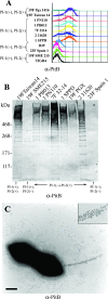
Detection of a functional surface-exposed pilus structure encoded by PI-2. (A) FACS analysis performed on S. pneumoniae clinical isolates (OD600 of 0.2) containing PI-1, PI-2, or both (presence or absence indicated by a plus or minus sign, respectively) labeled with mouse polyclonal PitB antiserum (secondary antibody was fluorescein isothiocyanate labeled). (B) Immunoblot analysis of different S. pneumoniae mutanolysin extracts reacted with mouse polyclonal anti-PitB antiserum. (C) Immunogold localization of PitB in pili of S. pneumoniae PN110 whole cells. Inset shows an enlarged portion of the pilus. α, anti.
FIG. 3.
FIG. 3.
Effect of PI-2 gene deletions on PI-2 polymerization in S. pneumoniae Serotype 1. (A) Western blot performed with anti-PitB antibodies on mutanolysin extracts of PN110 knockout isogenic mutants (Table 3). The PitB monomer is indicated. (B to G) Immunogold labeling with anti-PitB antibodies of whole-cell PN110 deletion mutants. Mutants PN110ΔPI2 (B), PN110ΔsipA (D), PN110ΔpitB (E), and PN110ΔsrtG1 (F) show lack of pilus formation. Panels C and G represent PN110ΔpitA and PN110ΔsrtG2, respectively. Bacteria were charged on Formvar carbon grids and immunogold decorated with mouse anti-PitB (gold particle size, 5 nm). Scale bar, 0.2 μm. α, anti.
FIG. 4.
FIG. 4.
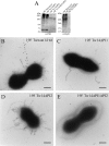
S. pneumoniae 19F Taiwan-14 strain expresses two independent pili. (A) Western blotting performed with polyclonal anti-PitB and anti-RrgB antibodies on mutanolysin extracts of 19F Taiwan-14 wild-type and knockout isogenic mutants lacking PI-1 and/or PI-2 (19F Tw14ΔPI1, Tw14ΔPI2, and Tw14ΔPI1ΔPI2). (B and E) Double immunogold labeling performed with mouse anti-PitB (gold particle size, 20 nm) and guinea pig anti-RrgB (gold particle size, 5 nm) on 19F Taiwan-14 wild type (wt) and 19F Tw14ΔPI1ΔPI2. (C) Deletion mutant 19F Tw14ΔPI1 labeled with mouse anti-PitB (gold particle size, 5 nm). (D) 19F Tw14ΔPI2 labeled with mouse anti-RrgB (gold particle size, 5 nm). Scale bar, 0.2 μm. α, anti.
FIG. 5.
FIG. 5.
(A1 to D2) Adherence assays of purified backbone protein to A549 cells. Cells grown on coverslips were incubated with 100 μg/ml of purified PitB (A1 and A2) or GFP (C1 and C2) in DMEM (B1 and B2) or with medium alone (D1 and D2) for 2 h at 4°C. Cells were labeled with anti-PitB (A1, A2, B1, and B2) and anti-GFP antibodies (C1, C2, D1, and D2) and phalloidin (A1, B1, C1, and D1). Imaging was performed with a confocal microscope. Scale bar, 10 μm. (E) Binding of purified proteins to A549 cells in suspension. The level of adherence was determined by FACS analysis at a range of protein doses (0, 5, 50, 100, and 200 μg/ml) of RrgA (triangles), RrgB (crosses), PitB (diamonds), and GFP (squares). Proteins were incubated for 2 h at 4°C, labeled with antiserum specific to each protein, and detected with Alexa Fluor 488-conjugated secondary antibodies. RrgA and GFP were used, respectively, as positive and negative controls. Cells were analyzed with a FACSCalibur flow cytometer, and the net MFI for each population was calculated from three independent experiments. Nonsignificant differences were detected between PitB, GFP, and RrgB (P > 0.05), and only RrgA was significantly different from GFP binding (P < 0.05). α, anti.
FIG. 6.
FIG. 6.
Cocultivation experiments. (A and B) A549 cells cocultivated with pneumococcal strains labeled with Omniserum primary antibodies and analyzed by confocal microscopy. (A) PN110 wild type. (B) PN110ΔPI2. (C) Confocal three-dimensional reconstruction of A549 cells cocultivated with PN110 wild type labeled with Omniserum and mouse anti-PitB antibodies. The arrow indicates the location of an extended pilus contacting host cells. In panels A to C, bacteria were visualized with Alexa Fluor 568-conjugated secondary antibodies (red), A549 cells were visualized with phalloidin conjugated to Alexa Fluor 647 secondary antibodies (blue), and PitB was visualized with Alexa Fluor 488-conjugated secondary antibodies (green). Scale bar, 10 μm. (D) Adherence quantification of different strains on A549 cells. (E) Adherence quantification of PN110 wild type and PN110ΔPI2 on various cell lines. For both panels D and E, bacteria were counted in five different microscope fields, and average results of three independent experiments are shown. Significant differences were detected by a Student's t test (*P < 0.05).

References

    1. Abbot, E. L., W. D. Smith, G. P. Siou, C. Chiriboga, R. J. Smith, J. A. Wilson, B. H. Hirst, and M. A. Kehoe. 2007. Pili mediate specific adhesion of Streptococcus pyogenes to human tonsil and skin. Cell Microbiol. 91822-1833. - "V体育官网" PubMed
    1. Aguiar, S. I., I. Serrano, F. R. Pinto, J. Melo-Cristino, and M. Ramirez. 2008. The presence of the pilus locus is a clonal property among pneumococcal invasive isolates. BMC Microbiol. 841. - PMC - PubMed
    1. Alloing, G., B. Martin, C. Granadel, and J. P. Claverys. 1998. Development of competence in Streptococcus pneumonaie: pheromone autoinduction and control of quorum sensing by the oligopeptide permease. Mol. Microbiol. 2975-83. - PubMed
    1. Barocchi, M. A., J. Ries, X. Zogaj, C. Hemsley, B. Albiger, A. Kanth, S. Dahlberg, J. Fernebro, M. Moschioni, V. Masignani, K. Hultenby, A. R. Taddei, K. Beiter, F. Wartha, A. von Euler, A. Covacci, D. W. Holden, S. Normark, R. Rappuoli, and B. Henriques-Normark. 2006. A pneumococcal pilus influences virulence and host inflammatory responses. Proc. Natl. Acad. Sci. USA 1032857-2862. - PMC - PubMed
    1. Basset, A., K. Trzcinski, C. Hermos, L. O'Brien, K., R. Reid, M. Santosham, A. J. McAdam, M. Lipsitch, and R. Malley. 2007. Association of the pneumococcal pilus with certain capsular serotypes but not with increased virulence. J. Clin. Microbiol. 451684-1689. - PMC (VSports最新版本) - PubMed

VSports - MeSH terms

Substances (VSports手机版)

"VSports" Associated data

"VSports app下载" LinkOut - more resources