Skip to main page content
U.S. flag

An official website of the United States government

Dot gov

The . gov means it’s official. Federal government websites often end in . gov or . mil VSports app下载. Before sharing sensitive information, make sure you’re on a federal government site. .

Https

The site is secure. The https:// ensures that you are connecting to the official website and that any information you provide is encrypted and transmitted securely V体育官网. .

. 2007 Dec;1(3):288-302.
doi: 10.1016/j.molonc.2007.10.003.

Vascular endothelial growth factor restores delayed tumor progression in tumors depleted of macrophages (VSports最新版本)

Affiliations

Vascular endothelial growth factor restores delayed tumor progression in tumors depleted of macrophages

Elaine Y Lin et al. Mol Oncol. 2007 Dec.

Abstract

Genetic depletion of macrophages in Polyoma Middle T oncoprotein (PyMT)-induced mammary tumors in mice delayed the angiogenic switch and the progression to malignancy. To determine whether vascular endothelial growth factor A (VEGF-A) produced by tumor-associated macrophages regulated the onset of the angiogenic switch, a genetic approach was used to restore expression of VEGF-A into tumors at the benign stages. This stimulated formation of a high-density vessel network and in macrophage-depleted mice, was followed by accelerated tumor progression. The expression of VEGF-A led to a massive infiltration into the tumor of leukocytes that were mostly macrophages VSports手机版. This study suggests that macrophage-produced VEGF regulates malignant progression through stimulating tumor angiogenesis, leukocytic infiltration and tumor cell invasion. .

Keywords: PyMT; VEGF; angiogenesis; macrophages; malignancy; mammary; mouse; progression; transgenic; tumor V体育安卓版. .


"V体育官网" Figures

Figure 1
Figure 1
Transgenic expression of VEGF in the mammary gland. (A) The expression of transgenic VEGF‐A and GFP was regulated by the tetracycline inducible system in which the expression of rtTA was under the control of the mammary‐specific promoter, MMTV‐LTR. Upon exposure to doxycycline (Dox), mRNA consisting of both VEGF‐A and GFP was synthesized in mammary epithelium. Subsequently, upon translation, 2A induced cleavage leads to the formation of the two distinct proteins, VEGF and GFP. (B) Western analysis of the transgenic expression. Western blots are prepared from mammary tissues of mice at 6weeks of age carrying the VEGF transgene, lanes 1 and 2 (lane 1, PyMT−; and lane 2, PyMT+) and control mice 3–5 (lane 3: PyMT+, rtTA−, VEGF+; lane 4: PyMT−, rtTA+, VEGF−; lane 5: PyMT+, rtTA−, VEGF+). Mice were treated with 1mg/ml Dox in the drinking water for 2weeks. The filter was probed with anti‐VEGF, ‐GFP and ‐β‐tubulin antibodies. (C) Tissue specific expression of the VEGF transgene. Western analysis of transgenic VEGF expression in various tissues as indicated. M. gland+, mammary gland from a VEGF transgenic mouse; M. gland−, mammary gland from a non‐transgenic control. β‐Tubulin was used as the loading control.
Figure 2
Figure 2
Transgenic expression of VEGF/GFP is mammary epithelium specific. Tissue sections prepared from Csf1r‐GFP and VEGF‐A bi‐transgenic mice. To mark the phagocytic macrophages, both mice were i.v. injected with Texas‐Red conjugated dextran 2h before the tissue isolation as described in Section 4. (a–d) Spleen sections from the Csf1r‐GFP (a and b) and the VEGF‐A bi‐transgenic (c and d) mice. The insets in a and c are shown at high magnification in b and d. Blue arrows point to the area consisting of mainly Texas‐Red dextran marked macrophages. Notice that in the spleen from Csf1r‐Gfp mice, most of the dextran‐labeled cells are also GFP‐positive (b). White arrows point to white pulp in the spleen which are the collections of lymphocytes. Notice that no GFP‐positive cells are in these areas. (e–h) Sections of thymus from the Csf1r‐Gfp (e and f) and the VEGF‐A bi‐transgenic (g and h) mice. The insets in e and g are shown at high magnification in f and h. Arrows point to cells that are dextran‐positive. Note that most of the dextran‐positive cells in Csf1r‐GFP thymus are also GFP‐positive (f). (i, j) Mammary gland sections from the Csf1r‐Gfp (i) and the VEGF‐A bi‐transgenic (j) mice. Arrows in j point to GFP‐positive mammary ducts and arrows in i point to GFP‐positive stromal cells. Scale bars for a, c, e, g, i, j: 100μm; for b, d, f, h: 20μm.
Figure 3
Figure 3
Transgenic expression of VEGF in the mammary gland induces vessel network formation. (A) Mammary whole mounts showing an increase of vessel density in the mammary gland isolated from a mouse carrying the VEGF transgene (ii) compared to a non‐transgenic control (i). (B) GFP expression in mouse mammary glands. Fluorescent micrograph of mammary whole mounts from a VEGF transgenic PyMT mouse (ii) and a non‐transgenic control (i) shows that both the mammary ducts and tumor lesions were marked by GFP. Bar: 1mm. (C) Transgenic VEGF induces vessel formation around the mammary ducts. The images were prepared from mammary whole mounts from a VEGF transgenic mouse i.v. injected with Texas‐Red dextran to label the blood vessels (ii). The mammary ducts are marked by the transgenic expression of GFP (i). The vessel network is formed surrounding the GFP expressing mammary ducts (iii). Bar: 0.3mm.
Figure 4
Figure 4
Transgenic expression of VEGF induces angiogenesis in pre‐malignant lesions. (A) Induction of a high density vessel network in pre‐malignant tumors from wild‐type PyMT mice. IHC using anti‐vWF antibodies. Section of mammary lesions from wild‐type PyMT mice at 6weeks of age carrying the VEGF transgene (right panel) or the non‐transgenic control (left panel) counter‐stained with hematoxylin. Mice were treated with Dox for 2weeks. The insets in the top panel are shown in the lower panel. The vessel lumen is labeled with an asterisk. Arrows in a and c point to vessel positively stained with the anti‐vWF antibody in the lesion. Scale bars for a and b: 100μm; for c and d: 30μm. (B) Induction of vessel network formation in adenoma stage tumors from Csf1op/Csf1op PyMT mice. (i) Sections of tumor lesions from Csf1op/Csf1op PyMT mice at 8weeks of age. Mice were treated with Dox for 2weeks. Blood vessels were marked by i.v. injection of Texas‐Red conjugated dextran and visualized by fluorescent microscopy. (ii) A tumor from VEGF transgenic mouse and (i), a non‐transgenic control. Arrows point to autofluorescent RBCs in capillaries. Bar: 30μm.
Figure 5
Figure 5
Transgenic expression of VEGF in mammary tumors accelerates tumor progression to malignancy. (A) H&E staining of tumor sections from Csf1op/Csf1op (a and b) and wild‐type (c and d) PyMT mice at 8weeks of age. Mice carrying the VEGF transgene: b and d; and the non‐transgenic controls: a and c. All of the mice were treated with 1μg/ml Dox for 2weeks. Bar: 100μm. (B) Quantitative analysis of tumor progression in VEGF transgenic and control PyMT mice. Genotypes: op‐non, Csf1op/Csf1op non‐transgenic mice; op trans, Csf1op/Csf1op mice carrying the MMTV‐rtTA/VEGF transgene; wt‐non, wild‐type non‐transgenic mice; wt trans, wild‐type mice carrying the MMTV‐rtTA/VEGF transgene. Statistical analysis: Fisher's Exact test.
Figure 6
Figure 6
Transgenic expression of VEGF in mammary glands induces leukocytic infiltration. (A) a–d, H&E staining of mammary ducts. b and d, mammary gland from a VEGF and MMTV‐rtTA double transgenic mouse; a and c, a non‐transgenic control. The insets in a and d are shown at high magnification in c and d. Notice that multiple layers of stromal cells are surrounding the mammary duct in the VEGF expressing transgenic mammary gland (d, arrows). Size bar for a and b: 100μm; for c and d: 30μm. (B) a–d, IHC of mammary lesions stained by an anti‐F4/80 antibody. b and d, mammary lesions from a VEGF transgenic mouse; a and c, lesions from a non‐MMV‐rtTA transgenic control. The insets in a and b are shown in c and d, respectively. RBC filled vessels are labeled by an asterisk. Scale bars for a and b: 100μm; for c and d: 30μm.
Figure 7
Figure 7
VEGF R1 and SDF‐1/CXCR4 expression in mammary tumors. (A) FACS analysis of VEGF R1 and CXCR4 expressing myeloid cells isolated from VEGF transgenic tumors. Myeloid cells isolated from mammary tumors of MMTV‐rtTA/VEGF transgenic mice were identified by FACS using anti‐CD11b and F4/80 antibodies (A, left). The expression of CXCR4 and VEGF receptor 1 in three different populations of myeloid cells, R2, R3 and R4, were further analyzed using FACS (A, right). (B) IHC staining of VEGF transgenic tumors for SDF1. A blood vessel is pointed out by short arrows (a) and positive stained cells in the tumor are pointed out by long arrows. Bar: 30μm.
Figure 8
Figure 8
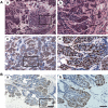
Transgenic expression of VEGF in Csf1op/Csf1op induces the formation of a “Medusa” structure. (A) Invasive tumor growth. (a, b) H&E staining of mammary tumors from a VEGF transgenic Csf1op/Csf1op PyMT mice at 8weeks of age. Black arrows in image a point to the invasive lesions and white arrows point to the “Medusa” structure. The invasive front of the tumor cells in the “Medusa” structure is shown in b, indicated by the arrows. (c, d) IHC using anti‐Ki67 antibody showing that a large percentage of tumor cells, especially these cells at the invasive front (arrows in d), in the “Medusa” structure is Ki67 positive. The insets in a and c are shown in b and d. (B) Ki67 IHC of mammary lesion from a non‐transgenic Csf1op/Csf1op mouse at 8weeks of age. The inset in a is shown in b. Notice that ∼50% of cells in the lesion are Ki67 negative. Scale bar for the left panel: 100μm; for the right panel: 30μm.

References

    1. Barleon, B. , Sozzani, S. , Zhou, D. , Weich, H.A. , Mantovani, A. , Marme, D. , 1996. Migration of human monocytes in response to vascular endothelial growth factor (VEGF) is mediated via the VEGF receptor flt-1. Blood. 87, 3336–3343. - PubMed (V体育安卓版)
    1. Cho, C.H. , Koh, Y.J. , Han, J. , Sung, H.K. , Jong Lee, H. , Morisada, T. , Schwendener, R.A. , Brekken, R.A. , Kang, G. , Oike, Y. , Choi, T.S. , Suda, T. , Yoo, O.J. , Koh, G.Y. , 2007. Angiogenic role of LYVE-1-positive macrophages in adipose tissue. Circ. Res.. 100, e47–e57. - PubMed (V体育官网)
    1. Dong, J. , Grunstein, J. , Tejada, M. , Peale, F. , Frantz, G. , Liang, W.C. , Bai, W. , Yu, L. , Kowalski, J. , Liang, X. , Fuh, G. , Gerber, H.P. , Ferrara, N. , 2004. VEGF-null cells require PDGFR alpha signaling-mediated stromal fibroblast recruitment for tumorigenesis. EMBO J. 23, 2800–2810. - PMC - PubMed
    1. Donnelly, M.L. , Hughes, L.E. , Luke, G. , Mendoza, H. , ten Dam, E. , Gani, D. , Ryan, M.D. , 2001. The ‘cleavage’ activities of foot-and-mouth disease virus 2A site-directed mutants and naturally occurring ‘2A-like’ sequences. J. Gen. Virol.. 82, 1027–1041. - PubMed
    1. Dvorak, H.F. , 2002. Vascular permeability factor/vascular endothelial growth factor: a critical cytokine in tumor angiogenesis and a potential target for diagnosis and therapy. J. Clin. Oncol.. 20, 4368–4380. - PubMed

Publication types

MeSH terms (V体育ios版)

Substances

"VSports" LinkOut - more resources