Skip to main page content
U.S. flag

An official website of the United States government

Dot gov

The . gov means it’s official. Federal government websites often end in . gov or . mil. Before sharing sensitive information, make sure you’re on a federal government site. VSports app下载.

Https

The site is secure. The https:// ensures that you are connecting to the official website and that any information you provide is encrypted and transmitted securely. V体育官网.

. 2008 Jun 3;105(22):7797-802.
doi: 10.1073/pnas.0800928105. Epub 2008 May 28.

T cell receptor signaling controls Foxp3 expression via PI3K, Akt, and mTOR

Affiliations

T cell receptor signaling controls Foxp3 expression via PI3K, Akt, and mTOR

Stephan Sauer et al. Proc Natl Acad Sci U S A. .

Abstract

Regulatory T (Treg) cells safeguard against autoimmunity and immune pathology. Because determinants of the Treg cell fate are not completely understood, we have delineated signaling events that control the de novo expression of Foxp3 in naive peripheral CD4 T cells and in thymocytes. We report that premature termination of TCR signaling and inibition of phosphatidyl inositol 3-kinase (PI3K) p110alpha, p110delta, protein kinase B (Akt), or mammalian target of rapamycin (mTOR) conferred Foxp3 expression and Treg-like gene expression profiles VSports手机版. Conversely, continued TCR signaling and constitutive PI3K/Akt/mTOR activity antagonised Foxp3 induction. At the chromatin level, di- and trimethylation of lysine 4 of histone H3 (H3K4me2 and -3) near the Foxp3 transcription start site (TSS) and within the 5' untranslated region (UTR) preceded active Foxp3 expression and, like Foxp3 inducibility, was lost upon continued TCR stimulation. These data demonstrate that the PI3K/Akt/mTOR signaling network regulates Foxp3 expression. .


Conflict of interest statement

The authors declare no conflict of interest.

Figures
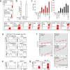
Fig. 1.
Fig. 1.
TCR signal deprivation and inhibitors of the PI3K/mTOR pathway induce Foxp3 expression in newly activated CD4 T cells. (a) Naive CD62LhiCD4+CD25 LN T cells were isolated by flow cytometry, labeled with CFSE, and activated for 18 h with anti-TCR and anti-CD28. After this, the cells were cultured for 36 h under various conditions: with plate-bound anti-TCR (with TCR signaling); without anti-TCR (no TCR signaling); and with the PI3K inhibitor LY294002 (LY, 10 μM), the mTOR inhibitor rapamycin (rapa, 25 nM), and/or TGFβ (1 ng/ml). Foxp3 expression was assessed by intracellular staining. (b) Naive CD4 T cells were activated for 18 h as in a and then cultured for 36 h with anti-TCR (with TCR signaling) or without anti-TCR (no TCR signaling). The expression of Foxp3 RNA was assessed by real time RT-PCR (mean ± SD, n = 3). (c) Naive CD4 T cells were activated for 18 h and aliquots of 105 cells (input) were transferred to the conditions described in a. After 36 h, cells were counted, and Foxp3 expression assessed by intracellular staining. Bars show the percentage (red) and the number (black) of Foxp3+ cells. (d) AND TCR transgenic Rag1−/− CD62LhiCD4+CD25 LN T cells were activated for 18 h as in a and then cultured for 36 h with anti-TCR (with TCR signaling); without anti-TCR (no TCR signaling); and with LY294002 (LY, 10 μM), rapamycin (rapa, 25 nM), or TGFβ (1 ng/ml). See Fig. S1 for percentages and numbers of Foxp3+ cells. (e) CD4+CD8CD25 thymocytes were activated for 18h as in a and then cultured for 36 h with anti-TCR (control) or with LY294002 and rapamycin without anti-TCR (LY+rapa). (f) Naive CD4 T cells were activated for 18 h as in a and cultured for 36 h with the indicated PI3K inhibitors without anti-TCR. The percentage of Foxp3+ cells is shown after subtracting Foxp3+ cells generated in cultures without anti-TCR (mean ± SD, n = 4–12). The p110 isoform specificity of each inhibitor is summarized in Table S1. (g) Naive CD4 T cells were activated for 18 h and cultured for 36 h with plate-bound anti-TCR (control) or with the allosteric Akt inhibitor Akti1/2 (0.3 μM) without anti-TCR.
Fig. 2.
Fig. 2.
TCR signaling controls the expression of Foxp3 via PI3K/mTOR/Akt. (a) Naive LN CD4 T cells were stimulated with anti-TCR and anti-CD28 for 1 or 18 h or for 1 h with anti CD28 alone (no TCR signaling). S6 phosphorylation was determined by intracellular staining. (b) Naive LN cells were activated as in a. After 18 h, rapamycin (25 nM) or LY294002 (10 μM) were added for 1 h, or the cells were cultured for the indicated time in the absence of anti-TCR. pS6 levels were determined as in a. (Inset) Immunoblotting confirmed declining pS6 in response to TCR signal deprivation, rapamycin, and LY294002. (c) (Top) PTEN-deficient and control CD4 T cells were depleted of preexisting Treg cells and activated with anti-TCR/CD28. (Middle) TGFβ (0.3 ng/ml) was added, and Foxp3 expression was assessed 48 h later. (Bottom) As a control, the PI3K inhibitor LY294002 was added and cells were deprived of TCR signaling after 18 h of activation.
Fig. 3.
Fig. 3.
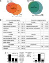
PI3K/mTOR inhibition initiates a Treg-like transcriptional program in newly activated T cells. (a) Gene expression differences between Treg cells versus naive CD4 T cells and in 24 h PI3K/mTOR inhibitor-treated cells versus naive CD4 T cells. Shown are the numbers of up- and down-regulated transcripts and the intersection of expression differences between Treg and Foxp3 induced cells. (b) Functional annotation of coregulated transcripts in Treg cells and Foxp3 induced cells relative to naive CD4 T cells, using DAVID. (c) microRNA expression by Treg cells and Foxp3 induced cells compared by qPCR. miR-142-3p and miR-21 are shown as examples for Treg-like and non-Treg-like microRNA expression by Foxp3 induced cells. The expression profiles of eight other microRNAs are listed.
Fig. 4.
Fig. 4.
No apparent TGFβ involvement in Foxp3 induction by PI3K and mTOR inhibitors. (a) Naive LN CD4 T cells were activated for 18 h in serum-free AIM-V medium and then exposed to TGFβ (1 ng/ml, 90 min, lane 1); TCR signal deprivation (90 min, lane 2); or TCR signal deprivation, rapamycin, and LY294002 (90 min for lane 3, 8 h for lane 4). Blots were sequentially probed with anti-pSmad2 (S465/467) and anti-Smad2/3. (b) Naive LN CD4 T cells activated as in a were deprived of TCR signals and TGFβ, and PI3K/mTOR inhibitors were added as indicated. Cultures were supplemented with neutralizing anti TGFβ (3 μg/ml) or the Smad kinase inhibitor SB431542. Foxp3 expression in the presence of anti TGFβ (dark gray bars) or SB431542 (light gray bars) was determined 2 days later and normalized to control cultures (black).
Fig. 5.
Fig. 5.
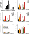
Inducibility of Foxp3 by PI3K and mTOR inhibition is transient, and H3K4 methylation at the Foxp3 TSS and 5′ UTR marks the inducible state. Naive CD4 T cells were activated for the indicated time with anti-TCR and anti-CD28 and then cultured for 36 h with LY294002 and rapamycin (Upper Left). Naive CD4 T cells (black), Treg cells (white), naive CD4 T cells activated for 18 h (orange) or 72 h (green) or induced to express Foxp3 by PI3K/mTOR inhibitors (red) were examined for H3K4me2 and H3K4me3 by ChIP and qPCR near the Foxp3 TSS (−295 to −105) and within the 5′ UTR (+4505 to +4621 and +6144 to +6280). Primer positions are indicated (34). pOct4 and pIkzf1 are shown as controls.

References

    1. Fisher AG. Cellular identity and lineage choice. Nat Rev Immunol. 2002;2:977–982. - PubMed
    1. Fontenot JD, Rudensky AY. A well adapted regulatory contrivance: Regulatory T cell development and the forkhead family transcription factor Foxp3. Nat Immunol. 2005;6:331–337. - PubMed
    1. Sakaguchi S. Naturally arising Foxp3-expressing CD25+CD4+ regulatory T cells in immunological tolerance to self and non-self. Nat Immunol. 2005;6:345–352. - PubMed
    1. Marie JC, Letterio JJ, Gavin M, Rudensky AY. TGF-β1 maintains suppressor function and Foxp3 expression in CD4+CD25+ regulatory T cells. J Exp Med. 2005;201:1061–1067. - PMC - PubMed
    1. Brunkow ME, et al. Disruption of a new forkhead/winged-helix protein, scurfin, results in the fatal lymphoproliferative disorder of the scurfy mouse. Nat Genet. 2001;27:68–73. - PubMed

Publication types

MeSH terms (V体育ios版)