"VSports手机版" Skip to main page content
U.S. flag

An official website of the United States government

Dot gov

The . gov means it’s official VSports app下载. Federal government websites often end in . gov or . mil. Before sharing sensitive information, make sure you’re on a federal government site. .

Https

The site is secure V体育官网. The https:// ensures that you are connecting to the official website and that any information you provide is encrypted and transmitted securely. .

. 2008 May 23;30(4):403-14.
doi: 10.1016/j.molcel.2008.03.009.

The CUL7 E3 ubiquitin ligase targets insulin receptor substrate 1 for ubiquitin-dependent degradation

Affiliations

V体育官网入口 - The CUL7 E3 ubiquitin ligase targets insulin receptor substrate 1 for ubiquitin-dependent degradation

Xinsong Xu et al. Mol Cell. .

V体育ios版 - Abstract

Recent genetic studies have documented a pivotal growth-regulatory role played by the Cullin 7 (CUL7) E3 ubiquitin ligase complex containing the Fbw8-substrate-targeting subunit, Skp1, and the ROC1 RING finger protein. In this report, we identified insulin receptor substrate 1 (IRS-1), a critical mediator of the insulin/insulin-like growth factor 1 signaling, as a proteolytic target of the CUL7 E3 ligase in a manner that depends on mammalian target of rapamycin and the p70 S6 kinase activities. Interestingly, while embryonic fibroblasts of Cul7-/- mice were found to accumulate IRS-1 and exhibit increased activation of IRS-1's downstream Akt and MEK/ERK pathways, these null cells grew poorly and displayed phenotypes reminiscent of those associated with oncogene-induced senescence. Taken together, our findings demonstrate a key role for the CUL7 E3 in targeting IRS-1 for degradation, a process that may contribute to the regulation of cellular senescence VSports手机版. .


"V体育ios版" Figures
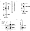
Figure 1
Figure 1. Analysis of Fbw8/IRS-1 Interaction In Vivo
(A and B) Analysis of the Flag-Fbw8 immunoprecipitates. Silver stain (A) and immunoblot (B) analysis of Flag-Fbw8-associated proteins, isolated as described in the Supplemental Data. (C) Interactions between ectopically expressed Fbw8 and IRS-1. HEK293 cells were transfected with vectors expressing V5-IRS-1 and Myc-Fbw8. At 48 hr posttransfection, cells were harvested. Extracts (500 µg) were used for immunoprecipitation with anti-Myc or anti-V5 antibodies followed by separation through 6% SDS-PAGE and immunoblot analysis as indicated. (D) Association between endogenous IRS-1 and CUL7. Extracts (1 mg of protein) from MCF-7 cells were subject to immunoprecipitation with IgG (lane 1) or anti-CUL7 (Mab38) antibodies (lane 2). The precipitates were separated by 6% SDS-PAGE followed by immunoblot analysis.
Figure 2
Figure 2. Fbw8 Promotes the Degradation of IRS-1
(A) Enhanced degradation of IRS-1 in HEK293 cells that overexpress Fbw8. HEK293 cells were cotransfected with vectors expressing V5-IRS-1, Myc-Fbw8, and HA-Ub, singly or in combination. At 42 hr posttransfection, where indicated, cells were treated with MG132 (10 µM) for 6 hr. Extracts (50 µg of protein) were subject to direct immunoblot analysis to monitor the changes in the level of V5-IRS-1 and to reveal the presence of Myc-Fbw8. Equal loading was verified by anti-HSP90 immunoblot. Note that the protein levels of β-actin (Figure 4B), HSP90, and Ku70 (Figure 2C and Figure 6A) were found unchanged in cells ectopically expressing Fbw8 and/or CUL7, and therefore each serves as an internal control for equal loading of samples in the immunoblot analysis described throughout the text. To reveal polyubiquitination, extracts (~1 mg of protein) from HEK293 cells were immunoprecipitated with anti-V5 antibodies. The precipitates were separated by 6% SDS-PAGE followed by immunoblot with anti-HA antibodies. (B) The specificity of the Fbw8-induced degradation of IRS-1. HEK293 cells were cotransfected with vectors expressing V5-IRS-1, Myc-Fbw8, or other F box proteins as indicated and were analyzed as in (A). The expression of Myc-Fbw8 or other F box proteins was determined by sequential immunoblot analysis, first reacted with anti-Myc and then anti-HA antibodies, followed by visualization using the Odyssey Infrared Imaging System. (C) CHX chase analysis. MCF-7 cells were transfected with pcDNA3.1 empty vectors (lanes 1–3) or vectors expressing Flag-CUL7 and Myc-Fbw8 (lanes 4–6). At 42 hr posttransfection, cells were treated with CHX (25 µg/ml) and chased for times as indicated. Extracts (50 µg) were separated by 6% SDS-PAGE followed by immunoblot analysis to monitor the levels of endogenous IRS-1, and ectopically expressed Flag-CUL7 and Myc-Fbw8. The remaining IRS-1 was quantitated by the Odyssey Infrared Imaging System and expressed graphically.
Figure 3
Figure 3. mTOR Is Required for the Degradation of IRS-1 by Fbw8
(A) CHX chase. MCF-7 cells were transfected with pcDNA3.1 empty vectors (lanes 1–4) or vectors expressing Flag-CUL7 and Myc-Fbw8 (lanes 5–12). At 24 hr posttransfection, cells were treated with (lanes 9–12) or without (lanes 1–8) rapamycin (100 nM). Twenty-four hours later, CHX (25 µg/ml) was added and chased for times as indicated, followed by immunoblot analysis as described in Figure 2C. (B) Enhanced degradation of IRS-1 by Fbw8 and Rheb. MCF-7 cells were cotransfected with vectors expressing Myc-Fbw8 and HA-Rheb. At 48 hr posttransfection, cells were harvested and the extracts (50 µg of protein) were analyzed by immunoblot to monitor the changes in the level of endogenous IRS-1 and to reveal the presence of ectopically expressed proteins. (C) A model for a role of the CUL7 E3 in the mTOR-IRS-1 negative-feedback regulation. Following insulin/IGF-1-mediated activation of receptors, IRS-1 becomes tyrosyl-phosphorylated and docks PI3-K, leading to activation of Akt and then Rheb, which turns on mTOR/S6K. The elevated mTOR/S6K serine-phosphorylate IRS-1, which sets the CUL7 E3-dependent ubiquitination and eventual degradation.
Figure 4
Figure 4. Analysis of IRS-1 Degradation Signals
(A) Elimination of phosphorylation at S307/S312/S527/S636/S639 renders V5-IRS-1 partially resistant to degradation by Fbw8. HEK293 cells were cotransfected with vectors expressing V5-IRS-1, Myc-Fbw8, or various point mutants as indicated and were analyzed as described in Figure 2A. 5A refers to a V5-IRS-1 quintuple mutant, replacing S307/S312/S527/S636/S639 with alanines. Of note, the wild-type V5-IRS-1 and variants V5-IRS-1S307A/S312A, V5-IRS-1S527A, and V5-IRS-1S636A/S639A appeared in hyperphosphorylated forms predominantly (lanes 16–19). In contrast, nearly half of V5-IRS-15A was present in hypophosphorylated species (lane 20), suggesting that the quintuple mutation significantly reduced hyperphosphorylation of IRS-1. These findings argued for a positive correlation between IRS-1 hyperphosphorylation and Fbw8-imposed degradation. (B) Overexpression of Fbw8 induces the degradation of IRS-1 (1–574) and IRS-1 (1–626). HEK293 cells were cotransfected with vectors expressing Myc-Fbw8, V5-IRS-1 (amino acids 1–1242), or its truncated derivatives as indicated and were analyzed as described in Figure 2A.
Figure 5
Figure 5. The CUL7 E3 Binds IRS-1 and Promotes Its Polyubiquitination In Vitro
(A) Dephosphorylation of IRS-1. Purified insect-cell-produced human IRS-1 (60 ng) was incubated with λ phosphatase (1 unit; lane 3) or alkaline phosphatase (1 unit; lane 5) at 37°C for 30 min. The reaction products were analyzed by immunoblot analysis using anti-IRS-1 antibody. (B) Phosphorylation of IRS-1 by S6K. Purified human IRS-1 (60 ng) was incubated with S6K (0.1, 1, and 10 pg for lanes 2–4, respectively) at 37°C for 30 min. The reaction products were analyzed by immunoblot analysis using antibodies recognizing IRS-1 phosphorylated at Ser307 (upper) or for total IRS-1. (C) In vitro interaction between Fbw8 and IRS-1. The binding experiment was carried out as described in the Supplemental Data, with input (20%) shown in lane 1. (D and E) Reconstitution of in vitro polyubiquitination of IRS-1 by the CUL7 E3 and kinetic analysis. Polyubiquitination of IRS-1 was reconstituted as described in the Experimental Procedures.
Figure 6
Figure 6. Inactivation of Fbw8 or CUL7 Accumulates IRS-1
(A) SiRNA-mediated depletion of Fbw8. FF8HEK cells, constitutively expressing Flag-Fbw8, were treated with scramble siRNA (lane 2) or Fbw8-siRNA (lane 3) at a concentration of 10 nM. SiRNA was obtained from Dharmacon, and transfection was carried out following manufacturers’ instruction. At 48 hr posttransfection, cells were harvested. Extracts (50 µg) were separated by 6% SDS-PAGE followed by immunoblot analysis to monitor the levels of Flag-Fbw8. (B) Fbw8 depletion accumulates IRS-1. MCF-7 cells were transfected with scramble or Fbw8 siRNA at indicated concentrations for 48 hr. Extracts (50 µg) were subject to immunoblot analysis to monitor the levels of endogenous IRS-1. (C) Accumulation of IRS-1 in Cul7−/− MEFs. MEFs were serum-starved for 16 hr prior to treatment with (lanes 2 and 4) or without (lanes 1 and 3) IGF-1 (3 nM) for 30 min. Extracts (100 µg of protein) were separated by 6% SDS-PAGE followed by immunoblot analysis to monitor the levels of endogenous IRS-1. (D) RT-PCR analysis of IRS-1 mRNA. IRS-1 mRNA from CUL7+/+ or CUL7−/− MEFs was analyzed using a procedure described in the Supplemental Data. Note that the graph integrates the results of three independent experiments, with error bars for the calculated standard deviation. (E–G) CUL7−/− MEFs accumulate IRS-1 and exhibit increased activation of Akt and MEK/ERK. MEFs derived from CUL7+/+ (lanes 1–6) or CUL7−/− (lanes 7–12) were serum-starved for 16 hr prior to treatment with (lanes 2–6 and 8–12) or without (lanes 1 and 7) IGF-1 (3 nM) for indicated times. Extracts (100 µg of protein) were separated by 4%–20% SDS-PAGE followed by immunoblot analysis to monitor the levels of CUL7, IRS-1, phosphorylated Akt, MEK, and ERK, as well as HSP90 (as a control for equal loading; note that CUL7+/+ and CUL7−/− MEFs contain identical levels of α-actinin, HSP90, or tubulin [Figure S6D]). The relative ratio of phosphorylated Atk (or ERK) versus total pool is quantitated and shown graphically. Note that in Figure 6E, lane 12, there was a reduction of IRS-1 at 120 min after IGF-1 treatment. This decrease, however, was not reproducible and is most likely an experimental variation.
Figure 7
Figure 7. CUL7−/− MEFs Show Senescence Phenotypes
(A) CUL7−/− MEFs are G1 arrested. Normal and CUL7−/− MEFs at passage 5 were analyzed by flow cytometry. Histograms of propidium iodide-stained cells are shown. The percentage of cells in G1, S, and G2/M phases of the cell cycle is indicated. (B) CUL7−/− MEFs induce p16 and exhibit hypophosphorylated pRb. Extracts (100 µg of protein) from normal or CUL7−/− MEFs at passage 5 were subject to immunoblot analysis to monitor the levels of p16, pRb, and α-actinin (as a control for equal loading). (C) CUL7−/− MEFs exhibited elevated levels of β-gal activity. Normal and CUL7−/− MEFs were stained for senescence-associated β-gal activity using a protocol as described (Serrano et al., 1997). Top and bottom panels show photographs of the normal and CUL7−/− MEFs at 100× and 400× magnification, respectively. β-gal-positive CUL7+/+ and CUL7−/− MEFs at passage 2 and 5 were quantitated and shown graphically. Note that the graph integrates the results of three independent experiments, with error bars for the calculated standard deviation. (D) Cell morphology of CUL7+/+ and CUL7−/− MEFs at passage 2 and 7. Morphology analysis was carried out as described in the Supplemental Data.

References

    1. Alessi DR, Cuenda A, Cohen P, Dudley DT, Saltiel AR. PD 098059 is a specific inhibitor of the activation of mitogen-activated protein kinase kinase in vitro and in vivo. J. Biol. Chem. 1995;270:27489–27494. - PubMed
    1. Andrews P, He YJ, Xiong Y. Cytoplasmic localized ubiquitin ligase cullin 7 binds to p53 and promotes cell growth by antagonizing p53 function. Oncogene. 2006;25:4534–4548. - PubMed
    1. Arai T, Kasper JS, Skaar JR, Ali SH, Takahashi C, DeCaprio JA. Targeted disruption of p185/Cul7 gene results in abnormal vascular morphogenesis. Proc. Natl. Acad. Sci. USA. 2003;100:9855–9860. - PMC - PubMed
    1. Campisi J. Senescent cells, tumor suppression, and organismal aging: good citizens, bad neighbors. Cell. 2005;120:513–522. - PubMed
    1. Daud AI, Lanson NA, Jr, Claycomb WC, Field LJ. Identification of SV40 large T-antigen-associated proteins in cardiomyocytes from transgenic mice. Am. J. Physiol. 1993;264:H1693–H1700. - "V体育2025版" PubMed

Publication types (V体育官网入口)

MeSH terms

Substances