VSports手机版 - Skip to main page content
U.S. flag

An official website of the United States government

Dot gov

The . gov means it’s official. Federal government websites often end in VSports app下载. gov or . mil. Before sharing sensitive information, make sure you’re on a federal government site. .

Https

The site is secure. The https:// ensures that you are connecting to the official website and that any information you provide is encrypted and transmitted securely V体育官网. .

. 2008 Jun;118(6):2098-110.
doi: 10.1172/JCI34584.

Tumor therapy in mice via antigen targeting to a novel, DC-restricted C-type lectin

Affiliations

Tumor therapy in mice via antigen targeting to a novel, DC-restricted C-type lectin (VSports在线直播)

David Sancho et al. J Clin Invest. 2008 Jun.

Abstract

The mouse CD8alpha+ DC subset excels at cross-presentation of antigen, which can elicit robust CTL responses. A receptor allowing specific antigen targeting to this subset and its equivalent in humans would therefore be useful for the induction of antitumor CTLs. Here, we have characterized a C-type lectin of the NK cell receptor group that we named DC, NK lectin group receptor-1 (DNGR-1). DNGR-1 was found to be expressed in mice at high levels by CD8+ DCs and at low levels by plasmacytoid DCs but not by other hematopoietic cells. Human DNGR-1 was also restricted in expression to a small subset of blood DCs that bear similarities to mouse CD8alpha+ DCs. The selective expression pattern and observed endocytic activity of DNGR-1 suggested that it could be used for antigen targeting to DCs. Consistent with this notion, antigen epitopes covalently coupled to an antibody specific for mouse DNGR-1 were selectively cross-presented by CD8alpha+ DCs in vivo and, when given with adjuvants, induced potent CTL responses. When the antigens corresponded to tumor-expressed peptides, treatment with the antibody conjugate and adjuvant could prevent development or mediate eradication of B16 melanoma lung pseudometastases. We conclude that DNGR-1 is a novel, highly specific marker of mouse and human DC subsets that can be exploited for CTL cross-priming and tumor therapy. VSports手机版.


"VSports app下载" Figures

Figure 1
Figure 1. Biochemical characterization of mouse DNGR-1 protein.
(A) Lysate from Phoenix cells stably transfected with a retroviral vector encoding HA-tagged mDNGR-1 was Western blotted (WB) with anti-HA before or after immunoprecipitation with anti-HA or the indicated anti–mDNGR-1 antibodies. (B) Western blot using anti–DNGR-1 mAb 397 to probe lysates of Phoenix cells expressing (+) or not expressing (–) DNGR-1 under reducing (right) and nonreducing (left) conditions.
Figure 2
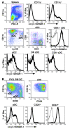
Figure 2. Mouse DNGR-1 expression is restricted to CD8α+ DCs and pDCs.
(A) Expression of DNGR-1 in mouse spleen. Splenocytes were stained with biotin anti–DNGR-1 7H11 (thick line) or biotin rat IgG1 (thin line; isotype-matched control) followed by streptavidin-PE and antibodies to CD11c, CD4, CD8α, and Ly6C. CD11c+ and CD11c cells (upper row) or different DC subsets (lower rows) were defined by electronic gating as shown on the dot plots. DNGR-1 expression is shown in the corresponding histograms. Numbers indicate percentage of events in gate. (B) Expression of DNGR-1 in Flt3L-BMDCs. Histograms show staining with anti–DNGR-1 (thick line) compared with isotype-matched control (thin line) in B220+ cells corresponding to pDCs, CD24hiCD11blo cells corresponding to CD8α+ DCs, and CD24loCD11bhi cells corresponding to CD8α DCs after gating as indicated. Numbers represent percentage of events in the indicated gate.
Figure 3
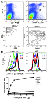
Figure 3. Specific labeling of CD8α+ DCs and pDCs in vivo with anti–DNGR-1 mAbs.
(A) Mice were injected i.v. with 100 μg of Alexa Fluor 488–conjugated anti–DNGR-1 (7H11) or isotype-matched control (rat IgG1), and splenocytes were analyzed 1 day later. Dot plots show CD11c versus Alexa Fluor 488 in anti–DNGR-1–injected (right panel) or rat IgG1–injected (left panel) mice. Contour plots show CD4 versus CD8α and Ly6C versus B220 profiles of the CD11chi and CD11cint DNGR-1+ populations circled in the dot plot. Numbers represent percentage of events in the indicated gate. (B) Mice were injected s.c. with the indicated doses of Alexa Fluor 488–conjugated anti–DNGR-1 (7H11) or a single dose of isotype-matched control (rat IgG1, 20 μg/mouse), and splenocytes were analyzed 1 day later. Histograms show staining of CD8α+ DCs or pDCs obtained with different doses of anti–DNGR-1 (green line, 0.5 μg; blue line, 2 μg; red line, 5 μg; thick black line, 20 μg) or isotype-matched control (thin black line). Lower panel shows MFI with anti–DNGR-1 divided by that obtained with 20 μg of isotype-matched control for each DC subset or for non-DC spleen cells.
Figure 4
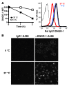
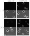
Figure 4. Endocytosis of anti–DNGR-1 mAbs in Flt3L BMDCs.
(A) Flt3L BMDCs were labeled with anti–DNGR-1 7H11 mAbs as detailed in Methods and incubated for the indicated times at 4°C or 37°C before adding streptavidin-PE. Data represent MFI for the CD24hi subset. Histograms show actual staining profiles: thick black line, staining with streptavidin-PE immediately after labeling with biotin–anti–DNGR-1; blue line, after 2 hours at 4°C; red line, after 2 hours at 37°C; thin black line, isotype-matched control. (B) Flt3L BMDCs were labeled with Alexa Fluor 488–conjugated anti–DNGR-1 mAbs (7H11, right panels) or isotype-matched control (rat IgG1, left panels) and incubated for 2 hours at 4°C (upper panels) or 37°C (lower panels) before confocal analysis. Image represents a single optical section (<0.7 μm). Note punctate staining inside cells incubated at 37°C, indicative of endocytosis. Original magnification, ×630.
Figure 5
Figure 5. Antigen targeted to DNGR-1 in vivo is cross-presented by CD8α+ DCs.
(A) 2 μg S1-conjugated anti–DNGR-1 (7H11) or rat IgG1 isotype-matched control mAbs were injected i.v. The next day, CD11c+ and CD11c spleen cell fractions were prepared, and graded numbers were cultured for 4 days with CFSE-labeled OT-I cells. Histograms show CFSE profiles of OT-I cells incubated with 25 × 103 CD11c or CD11c+ splenocytes. Right panels show absolute number of OT-I cells per well and IFN-γ content in supernatants. (B) The indicated DC subsets purified from mice injected with 2 μg S1–anti–DNGR-1 (7H11) were cultured for 3 days with CFSE-labeled OT-I cells. Response was analyzed as in A. Data are representative of at least 3 experiments.
Figure 6
Figure 6. CTL priming with antigen targeting to DNGR-1 plus anti-CD40.
2 μg S1-conjugated anti–DNGR-1 (7H11) or rat IgG1 isotype-matched control mAbs were injected s.c. with or without anti-CD40 (25 μg) as indicated. Target cells were injected 5 days later, and mice were analyzed on day 6. (A) In vivo CTL activity as measured by target cell elimination. Histograms show target cell frequency in representative mice from each group (CFSElo, 20 nM peptide; CFSEint, 200 nM peptide; CFSEhi, no peptide). Graph shows mean ± SEM of percentage of specific lysis in 1 experiment of 3 (n = 6 mice/group). All groups are shown, but the only one in which killing was detectable was that receiving anti–DNGR-1–S1 plus anti-CD40. (B) H-2Kb–SIINFEKL tetramer staining of splenocytes. Left panel shows representative dot plots of tetramer staining versus CD8 in gated CD8+ Thy1+ T cells. Right panel shows frequency of tetramer-positive CD8+ T cells in 1 experiment of 3 (n = 6 mice/group). (C) In vitro restimulation with 1 μM SIINFEKL (PEPTIDE) or medium alone (CTR). Left panel IFN-γ content in supernatants at the end of the 5-day culture. Right panel shows specific CTL activity of in vitro–restimulated cells against EL4 targets loaded with 2 μM of SIINFEKL. Data are the average + SEM of all cultures (n = 6 mice/group, restimulated individually). P values were calculated using Student’s t test. (D) OVA protein conjugated to anti–DNGR-1 induces CTL priming in vivo. Mice were immunized s.c. in the paw with 2 μg anti–DNGR-1 or isotype control antibodies conjugated to OVA protein in the presence of 25 μg of anti-CD40. In vivo killing activity was analyzed as in A using targets loaded with 200 nM SIINFEKL peptide. Results represent individual mice and the mean for 1 representative experiment out of 3. n = 5; P < 0.01, t test.
Figure 7
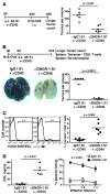
Figure 7. Anti–DNGR-1–S1 plus anti-CD40 is effective in prevention and therapy of OVA-expressing B16 melanoma.
(A) Tumor prevention experiments were carried out as depicted. Data represent lung tumor counts in each mouse in 1 experiment out of 2 (n = 6 mice/group). (B) Tumor therapy experiments were carried out as depicted. Lower left panel shows representative photographs of lungs from mice treated as indicated. Right panel shows quantification of tumors in each mouse in 1 representative experiment out of 3 (n = 6 mice/group). (C) SIINFEKL-H2Kb tetramer-positive cells in spleens from mice depicted in B. Left panels show representative histograms of gated CD8+Thy1.2+ splenocytes. Right panel shows frequency of tetramer-positive CD8+ T cells in 1 representative experiment out of 3 (n = 6 mice/group). (D) Left panel shows splenocytes from individual mice depicted in B restimulated in vitro with SIINFEKL peptide (1 μM). IFN-γ levels after 5 days of culture are indicated for 1 representative experiment out of 3 (n = 6 mice/group). Right panel shows CTL activity after restimulation measured as in Figure 6C. One experiment (n = 6 mice/group, restimulated individually) out of 3 is shown. Data are the average ± SEM of all cultures. P values were calculated using Student’s t test.
Figure 8
Figure 8. Immunotherapy of B16 melanoma via targeting of tumor antigens to DNGR-1.
(A) Tumor therapy experiments were carried out as depicted (top panel) using peptides encompassing known epitopes of melanocyte differentiation endogenous antigens (Endo: gp100, TRP-1, and TRP-2) covalently coupled to anti–DNGR-1 or to an isotype-matched control antibody. Poly I:C plus anti-CD40 was used as adjuvant. Lower left panel shows representative photographs of lungs from mice treated as indicated. Right panel shows quantification of lung tumors in each mouse. Data are pooled from 2 independent experiments (n = 9 mice/group), and each point represents 1 mouse. (B) Splenocytes from individual mice depicted in A were restimulated in vitro with the melanocyte differentiation antigen peptides used for immunization (10 μM). IFN-γ levels after 2 days of culture are shown. Data are pooled from 2 independent experiments (n = 9 mice/group). P values were calculated using the Mann-Whitney U test.
Figure 9
Figure 9. Human DNGR-1 expression is restricted to BDCA-3+ blood DCs.
(A) Human PBMCs were stained with anti–hDNGR-1 (8F9) or an isotype-matched control antibody (mouse IgG2a) and counterstained for various leukocyte markers. Histograms show DNGR-1 staining on T cells, B cells, monocytes, NK cells, lineage-negative HLA-DR cells, and lineage-negative HLA-DR+ cells. Numbers indicate the percentage of hDNGR-1+ cells in the latter fraction. (B) PBMCs depicted in A were gated on lineage-negative HLA-DR+ cells. Dot plots show staining with anti–DNGR-1 or isotype-matched control mAbs against various blood DC subset markers. Numbers represent percentage of cells in each quadrant.
Figure 10
Figure 10. Endocytosis of anti-human DNGR-1 mAbs by BDCA-3+ blood DCs.
Human blood BDCA-3+ DCs were stained with anti-hDNGR-1 (8F9) or an isotype-matched control antibody (mouse IgG2a), washed, and left at 4°C (upper panels) or 37°C (lower panels) for 2 hours before fixation and mounting. Representative confocal optical sections (<0.7 μm) are shown. Original magnification, ×1260.

References

    1. Steinman R. Dendritic cells: versatile controllers of the immune system. Nat. Med. 2007;13:1155–1159. doi: 10.1038/nm1643. - "VSports最新版本" DOI - PubMed
    1. Villadangos J., Schnorrer P. Intrinsic and cooperative antigen-presenting functions of dendritic-cell subsets in vivo. Nat. Rev. Immunol. 2007;7:543–555. doi: 10.1038/nri2103. - DOI - PubMed
    1. Vremec D., Shortman K. Dendritic cell subtypes in mouse lymphoid organs: cross-correlation of surface markers, changes with incubation, and differences among thymus, spleen, and lymph nodes. J. Immunol. 1997;159:565–573. - PubMed
    1. Henri S., et al. The dendritic cell populations of mouse lymph nodes. J. Immunol. 2001;167:741–748. - VSports注册入口 - PubMed
    1. den Haan J.M., Lehar S.M., Bevan M.J. CD8(+) but Not CD8(-) Dendritic cells cross-prime cytotoxic T cells in vivo. J. Exp. Med. 2000;192:1685–1696. doi: 10.1084/jem.192.12.1685. - DOI - PMC - PubMed

Publication types

MeSH terms