Skip to main page content
U.S. flag

An official website of the United States government

Dot gov

The . gov means it’s official VSports app下载. Federal government websites often end in . gov or . mil. Before sharing sensitive information, make sure you’re on a federal government site. .

Https

The site is secure V体育官网. The https:// ensures that you are connecting to the official website and that any information you provide is encrypted and transmitted securely. .

. 2008 Jun;14(6):633-40.
doi: 10.1038/nm1770. Epub 2008 May 18.

Persistent activation of an innate immune response translates respiratory viral infection into chronic lung disease

Affiliations

"V体育官网入口" Persistent activation of an innate immune response translates respiratory viral infection into chronic lung disease

Edy Y Kim (VSports在线直播) et al. Nat Med. 2008 Jun.

Abstract (V体育平台登录)

To understand the pathogenesis of chronic inflammatory disease, we analyzed an experimental mouse model of chronic lung disease with pathology that resembles asthma and chronic obstructive pulmonary disease (COPD) in humans. In this model, chronic lung disease develops after an infection with a common type of respiratory virus is cleared to only trace levels of noninfectious virus. Chronic inflammatory disease is generally thought to depend on an altered adaptive immune response. However, here we find that this type of disease arises independently of an adaptive immune response and is driven instead by interleukin-13 produced by macrophages that have been stimulated by CD1d-dependent T cell receptor-invariant natural killer T (NKT) cells. This innate immune axis is also activated in the lungs of humans with chronic airway disease due to asthma or COPD VSports手机版. These findings provide new insight into the pathogenesis of chronic inflammatory disease with the discovery that the transition from respiratory viral infection into chronic lung disease requires persistent activation of a previously undescribed NKT cell-macrophage innate immune axis. .


VSports最新版本 - Figures
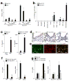
Figure 1
Figure 1
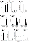
IL-13-producing macrophages drive chronic airway disease after viral infection. (a) Wild-type (WT) and Il13−/− mice were inoculated with SeV or UV-inactivated SeV (SeV-UV), and WT mice were untreated (NT) or treated with control IgG or sIL-13Rα2-Fc from Day 12 to 49 PI. Lung levels of Muc5ac mRNA were determined at Day 21 and 49 PI. (*) indicates a significant decrease from corresponding untreated control. (b) Lung cell subsets were isolated at Day 21 and 49 PI and were analyzed for Il-13 mRNA levels. Values represent Il-13 mRNA per cell fraction. (c) Lung CD4+ T cells and macrophages from Day 49 PI were cultured without or with PMA (20 ng/ml) and ionomycin (500 ng/ml) for 24 h at 37 °C, and IL-13 was determined in cell culture media. (*) indicates a significant difference from corresponding SeV-UV control. (d) Airway sections from WT or Il13−/− mice at Day 49 PI were immunostained for IL-13. (e) Airway sections from WT mice at Day 49 PI were immunostained for IL-13 and CD68. Bars = 20 μm. (f) Op/op mice at Day 49 PI were assessed for lung Il-13 and Muc5ac mRNA levels. (*) indicates a significant decrease from corresponding SeV-UV control value. (g) WT mice were assessed after being treated with clodronate (+) or empty control (−) liposomes from Day 12 to 49 PI. (*) indicates a significant decrease from corresponding value for empty liposome treatment.
Figure 2
Figure 2
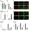
Activated NKT cells are required for chronic IL-13-producing macrophages in the lung after viral infection. (a) CD4+ and CD4 NKT cells were purified from mouse lungs at Day 0, 21, and 49 PI and were analyzed for Il-13 mRNA levels. Values represent units of Il-13 mRNA per cell fraction. (b) Corresponding number of cells in each NKT cell fraction. (c) Corresponding values for units of Il-13 mRNA per cell. For (a–c), (*) indicates a significant difference from Day 0 PI. (d) WT and Cd1d1−/− mice were inoculated with SeV or SeV-UV, and lung sections were immunostained for IL-13+CD68+ cells. (e) Using conditions in (d), mouse lungs were analyzed for Il-13 and Muc5ac mRNA levels. (f) WT and Traj18−/− mice were inoculated with SeV or SeV-UV, and lungs at Day 49 PI were analyzed for Il-13 and Muc5ac mRNA levels. (g). WT and Cd1d1−/− mice were inoculated with SeV or SeV-UV, and lung sections at Day 49 PI were immunostained for CD68+ cells. For (d–g), (*) indicates a significant decrease from corresponding value for WT mice. (h) CD4+ and CD4 NKT cells were purified from mouse lungs at PI Day 49 and were analyzed for Ccl3 mRNA by real-time PCR. (*) indicates a significant increase from SeV-UV.
Figure 3
Figure 3
Direct lung NKT cell-macrophage interaction generates IL-13-producing macrophages. (a) Lung macrophages were cultured alone or with lung NKT cells or CD4+ T cells for 24 h at 37 °C. Lung macrophages were also treated with anti-CD1d mAb or control IgG2b. (b) Culture conditions are the same as (a), but also include stimulation using PMA-ionomycin or α-GalCer (αGC). (c) Conditions are the same as (b), but macrophages were cultured with CD4+ and CD4 liver NKT cells. For (a–c), adherent macrophages were analyzed for Il-13 mRNA, and cell media was analyzed for corresponding IL-13 protein levels. Note 10–20-fold increase in y-axis for (b,c) compared to (a). (*) indicates a significant increase from macrophage and NKT cell alone, and (**) indicates a significant decrease from corresponding value for lung NTK cell-macrophage co-culture.
Figure 4
Figure 4
NKT cells drive IL-13R expression to upregulate IL-13 production and alternative activation of macrophages after viral infection. (a) Lung levels of Il-13ra1 mRNA in WT, Cd1d1−/−, and Il13−/− mice at Day 49 PI. (b) Levels of Il-13ra1 mRNA in macrophages (Mac1+ and CD3 NK1.1GR1B220CD11c) and non-macrophages (Mac1) purified from WT and Cd1d1−/− mice at Day 49 PI. Similar results were obtained for Mac1+CD68+ versus Mac1+CD68 cell subsets (data not shown). For (a,b), (*) indicates a significant increase from SeV-UV. (c) Representative photomicrographs of lungs sections immunostained for CD68 and Il-13rα1 at Day 49 PI. Bar = 50 μm. (d) Quantitative analysis of IL-13+CD68+ macrophages in mice inoculated with SeV-UV or SeV and treated with either control IgG or sIL-13Rα2-Fc from Day 12 to 49 PI. (e) Lung levels of Il-13 mRNA determined for conditions in (d). For (d,e), (*) indicates significant decrease from control IgG. (f) Representative photomicrographs of lungs sections immunostained for CD68 and Chi3l3/4 at Day 49 PI. Bar = 50 μm. (g) Lung levels of Chi3l3/4 mRNA in WT, Il13−/−, and Cd1d1−/−mice at Days 21 and 49 PI. (h) Levels of Chi3l3/4, Arg1, and Mmp12 mRNA in lung macrophages purified from WT and Cd1d1−/− mice at Day 49 PI. For (g,h), (*) indicates a significant increase from corresponding SeV-UV value, and (**) indicates a significant decrease from corresponding WT control value.
Figure 5
Figure 5
Increased IL-13-producing macrophages and Vα24-expressing NKT cells in humans with COPD and mucous cell metaplasia. (a) Representative photomicrographs of lung sections from a COPD subject undergoing lung transplantation and a non-COPD lung transplant donor were immunostained for MUC5AC. (b) Levels of MUC5AC and IL-13 mRNA in lungs from COPD subjects and non-COPD controls. (c) Representative photomicrographs of lung sections from COPD and non-COPD subjects that were immunostained for IL-13 and CD68. Arrows indicate an IL-13+CD68+ macrophage; arrowheads indicate an IL-13CD68+ macrophage. (d) Quantitative analysis of data from (c) in lungs from COPD subjects and non-COPD controls. (e) Representative photomicrographs of lung sections from COPD subjects that were immunostained for Vα24. Arrows indicate Vα24+ NKT cells. (f) Quantitative analysis of data from (e) in COPD subjects and non-COPD lung transplant donors. All bars = 50 μm. For (b,d,f), n = 5 per group, and (*) indicates a significant increase from non-COPD control value.
Figure 6
Figure 6
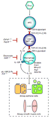
Scheme for an NKT cell-macrophage immune axis leading to chronic airway disease after viral infection. Virus may directly or indirectly facilitate CD1d-dependent antigen presentation and consequent activation of invariant CD4 NKT cells. NKT cells then interact directly with lung macrophages via contact between invariant Vα14 TCR and glycolipid-loaded CD1d. This interaction leads to increased expression of IL-13R and production of IL-13 that drives a positive feedback loop to amplify IL-13 production and alternative activation of macrophages, including Chi3l3/4, Fizz1, Mmp12, Arg1, and Alox12e gene expression. Persistent IL-13 production also drives differentiation of airway epithelial cell precursors towards mucous cells (mucous cell metaplasia) and airway smooth muscle cells to become more reactive to contractile agonists (airway hyperreactivity or AHR).

Comment in

References

    1. Mattner J, et al. Exogenous and endogenous glycolipid antigens activate NKT cells during microbial infections. Nature. 2005;434:525–529. - PubMed
    1. Seino K, Taniguchi M. Functional roles of NKT cells in the immune system. Frontiers Biosci. 2006;9:2577–2587. - PubMed
    1. Busse WW, Lemanske RFJ. Asthma. New Engl J Med. 2001;344:350–362. - PubMed
    1. Herrick CA, Bottomly K. To respond or not to respond: T cells in allergic asthma. Nat Rev Immunol. 2003;3:1–8. - PubMed
    1. Jones EY, Fugger L, Strominger JL, Siebold C. MHC Class II proteins and disease: a structural perspective. Nat Rev Immunol. 2006;6:271–282. - PubMed

V体育官网入口 - Publication types

MeSH terms

"V体育安卓版" Associated data