Skip to main page content
U.S. flag

An official website of the United States government

Dot gov

The . gov means it’s official. Federal government websites often end in . gov or VSports app下载. mil. Before sharing sensitive information, make sure you’re on a federal government site. .

Https

The site is secure V体育官网. The https:// ensures that you are connecting to the official website and that any information you provide is encrypted and transmitted securely. .

. 2008 Jun 9;205(6):1395-408.
doi: 10.1084/jem.20080277. Epub 2008 May 12.

V体育ios版 - Control of hematopoietic stem cell quiescence by the E3 ubiquitin ligase Fbw7

Affiliations

Control of hematopoietic stem cell quiescence by the E3 ubiquitin ligase Fbw7

Benjamin J Thompson et al. J Exp Med. .

Abstract

Ubiquitination is a posttranslational mechanism that controls diverse cellular processes. We focus here on the ubiquitin ligase Fbw7, a recently identified hematopoietic tumor suppressor that can target for degradation several important oncogenes, including Notch1, c-Myc, and cyclin E. We have generated conditional Fbw7 knockout animals and inactivated the gene in hematopoietic stem cells (HSCs), progenitors, and their differentiated progeny. Deletion of Fbw7 specifically and rapidly affects hematopoiesis in a cell-autonomous manner. Fbw7(-/-) HSCs show defective maintenance of quiescence, leading to impaired self-renewal and a severe loss of competitive repopulating capacity. Furthermore, Fbw7(-/-) progenitors are unable to colonize the thymus, leading to a profound depletion of T cell progenitors. Deletion of Fbw7 in bone marrow (BM) stem cells and progenitors leads to the stabilization of c-Myc, a transcription factor previously implicated in HSC self-renewal VSports手机版. On the other hand, neither Notch1 nor cyclin E is visibly stabilized in the BM of Fbw7-deficient mice. Gene expression studies of Fbw7(-/-) HSCs and hematopoietic progenitors indicate that Fbw7 regulates, through the regulation of HSC cycle entry, the transcriptional "signature" that is associated with the quiescent, self-renewing HSC phenotype. .


Figures
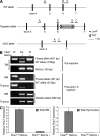
Figure 1.
Figure 1.
Generation of a conditional Fbw7 allele. (A) Targeting strategy showing exons 4–7 of the mouse genomic Fbw7 locus before targeting (WT allele) and after homologous recombination in C57BL/6 × 129 ES cells (targeted allele), which introduces an FRT- and LoxP-flanked PGK-Neo cassette between exons 6 and 7, and flanks exons 5 and 6 with LoxP sites. Germline removal of the PGK-Neo cassette using the FLPe-deleter strain and subsequent exposure to Cre recombinase results in excision of exons 5 and 6 and generates the conditional knockout (CKO) allele. Arrows show locations of primers (A–D) used to genotype resulting mice. (B) Before crossing to the FLPe-deleter strain, PCR using primers C and D were used to confirm transmission of the targeted allele (not depicted). After removal of the PGK-Neo cassette, primers B and C were used to detect both the “floxed” and WT alleles (497 and 315 bp, respectively). After induction of Mx-Cre with polyI-polyC injections, the floxed band was lost from the BM and thymus, and excision of exons 5 and 6 in these tissues was confirmed by PCR using primers A and C, which amplify 662 bp from the CKO allele. (C) Loss of WT Fbw7 expression was confirmed by quantitative RT-PCR using the same primers as in panel A, which are located in exons 4 and 5. Error bars show the SD of duplicate wells.
Figure 2.
Figure 2.
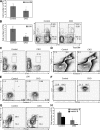
Fbw7 deletion leads to a depletion of BM hematopoietic progenitors. (A) Absolute numbers of BM cells in polyI-polyC–injected control (Fbw7f/f, MxCre) and CKO (Fbw7f/f, MxCre+) mice 2 wk after polyI-polyC deletion are shown. (B) Lin BM cells were stained for c-kit and Sca-1 to mark the LSK population (dot plot), and absolute LSK numbers (histogram) were calculated (also at 2 wk after polyI-polyC). (C) Quantitation of peripheral (spleen) LSKs 2 wk after polyI-polyC injection. P < 0.05. (D and E) Absence of enhanced rates of apoptosis as defined by a combination of 7-AAD/annexin V staining in either total BM of LSK cells (3 wk after polyI-polyC injection). (F) LSK cells were analyzed by FACS for CD34 expression to separate long-term (LT, CD34) from short-term (ST, CD34+) HSCs. (G) MPs (Lin/Sca-1/c-kit+) were analyzed by FACS for CD34 and FcγRII/III expression to show common MPs (CMP, CD34+/ FcγRII/III; bottom right gate), granulocyte/macrophage progenitors (GMP, CD34+/ FcγRII/III+; top right gate), and megakaryocyte/erythroid progenitors (MEP, CD34lo / FcγRII/III; bottom left gate). (H) Absolute number of mature (IgM+/B220hi) and immature (IgM+/B220+) BM B cells are shown (2 wk after polyI-polyC). Error bars show the SD. Numbers indicate the percentage of cells in each gate. *, P < 0.05; **, P < 0.005. For all experiments, n = 8.
Figure 3.
Figure 3.
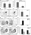
Loss of Fbw7 severely impairs early T cell development. (A) Absolute numbers of thymocytes (left, 2 wk after polyI-polyC; right, 4 wk after polyI-polyC) were counted from control (n = 8) and CKO (n = 8) mice and analyzed by FACS for CD4 and CD8 expression (B). (C) Absolute numbers of CD48 DN cells (2 wk after polyI-polyC). (D) DN cells were separated into DN1–4 populations by staining for CD44 and CD25. (E) Absolute numbers of DN2 (CD44+/CD25+) and DN3 (CD44/CD25+) (2 wk after polyI-polyC). (F) DN thymocytes were analyzed as in D in control (Fbw7f/f, LckCre) and CKO (Fbw7f/f, LckCre+) mice. Error bars show the SD. Numbers indicate the percentage of cells in each gate. **, P < 0.005. n = 6. (G) Absolute numbers of ETP (LinCD25CD44+c-kit+) cells 2 and 4 wk after polyI-polyC injection. *, P < 0.05. n = 4.
Figure 4.
Figure 4.
Loss of Fbw7 leads to a cell-autonomous defect of HSC self-renewal. (A) 0.5 × 10⁶ WT CD45.1 BM cells were injected into lethally irradiated WT Ly 5.1+ host mice (n = 3 for each experiment) with the same number of either control CD45.2 or CKO (Mx1-cre+Fbw7f/f) CD45.2 BM cells. The mice were analyzed 5 wk later for reconstitution of the BM, thymus, and spleen by staining cells from each tissue for both CD45.1 and CD45.2. The percent contribution of each donor to those organs is shown. CD45.2-gated cells from recipient mice were analyzed by FACS for expression of CD4, CD8, CD44, and CD25 in the thymus (D and E), B220/IgM (C), and LSK markers (B) in the BM. The cells in E are gated on the thymic Lin population. Numbers indicate the percentage of cells in each gate.
Figure 5.
Figure 5.
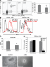
Deletion of Fbw7 leads to defective HSC quiescence and self-renewal. (A) LSK control and Mx1-cre+Fbw7f/f (CKO) mice were further analyzed for DNA content (DAPI) and intracellular Ki-67 expression (2 wk after polyI-polyC). Quadrants correspond to cell cycle stages as shown in the left panel. The percentage of LSK cells in G0 stage is also shown (n = 6), (B) BrdU labeling of the CD34 and CD34+ LSK subsets (n = 4) (2 wk after polyI-polyC). (C) 25,000 total BM cells from control or CKO mice were plated in methylcelluose medium containing a multilineage cytokine cocktail. The number of colonies and their qualitative morphologies were determined 8 d later. (D) The same experiment as in C was performed, except plating 500 flow-purified LT-HSCs per plate. After 7 d, the colonies were counted (first plating). The colonies from each plate were pooled and resuspended in PBS, and 2,000 of those cells were replated for an additional 7 d before counting colonies again (second plating). Numbers indicate the percentage of cells in each gate. (E) Photographs of representative colonies from C (first plating) show the relative size difference between control- and CKO-derived colonies. Bar, 0.5 mm. These are representatives of at least three individual experiments. *, P < 0.05; **, P < 0.005.
Figure 6.
Figure 6.
Effects of Fbw7 deletion on target protein stabilization in the BM. (A) Western blot detecting expression of Notch1-IC, uncleaved membrane Notch1 (Notch1-TM), c-Myc, cyclin E, and b-actin in total BM extracts. (B) Expression of c-Myc in cytokine-stimulated BM progenitors. LineageIL-7RαSca-1highc-kithigh cells from control and CKO (Mx-cre+Fbw7flox/flox) mice were cultured for 48 h in the presence of cytokines. c-Myc expression was quantified using densitometry and actin normalization. Phospho–c-Myc (T58/S62) and actin levels are also shown. A representative of three experiments is shown. (C) Quantitative RT-PCR quantifying expression of Fbw7 and c-Myc in the cells used in B. Expression is normalized using b-actin.
Figure 7.
Figure 7.
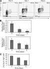
Dynamic regulation of Fbw7 expression during HSC cycle entry and differentiation. (A) Flow-purified LSK cells were cultured with cytokines for 0, 1, or 2 d and were stained for intracellular Ki-67 and DNA content (DAPI). (B) Fbw7 and Cul1 mRNA expression were analyzed by quantitative RT-PCR on each day. (C) Quantification of Sca-1 expression (loss of Sca-1 indicates differentiation of LSK cells) after each day of culture. On day 0, all cells are Sca-1+.
Figure 8.
Figure 8.
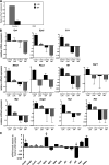
Fbw7 controls genes that correlate with HSC quiescence and self-renewal. (A) Efficient silencing of Fbw7 expression in LSK cells and MPs in response to polyI-polyC treatment (at 2 wk after treatment). (B) Quantitative RT-PCR validation of genes selected from the microarray using mRNA from a different sorting experiment. The plotted expression values represent the relative mRNA level of each sample normalized to the median expression across all samples. (C) Quantitative RT-PCR analysis of the expression of multiple cell cycle–related genes in control (Mx-cre+Fbw7+/+) and CKO (Mx-cre+Fbw7fl/fl) LSK cells.

References (VSports注册入口)

    1. Passegue, E., and A.J. Wagers. 2006. Regulating quiescence: new insights into hematopoietic stem cell biology. Dev. Cell. 10:415–417. - VSports - PubMed
    1. Aguila, H.L., K. Akashi, J. Domen, K.L. Gandy, E. Lagasse, R.E. Mebius, S.J. Morrison, J. Shizuru, S. Strober, N. Uchida, et al. 1997. From stem cells to lymphocytes: biology and transplantation. Immunol. Rev. 157:13–40. - "VSports最新版本" PubMed
    1. Adams, G.B., and D.T. Scadden. 2006. The hematopoietic stem cell in its place. Nat. Immunol. 7:333–337. - PubMed
    1. Yin, T., and L. Li. 2006. The stem cell niches in bone. J. Clin. Invest. 116:1195–1201. - PMC - PubMed
    1. Trowbridge, J.J., M.P. Scott, and M. Bhatia. 2006. Hedgehog modulates cell cycle regulators in stem cells to control hematopoietic regeneration. Proc. Natl. Acad. Sci. USA. 103:14134–14139. - PMC - PubMed

Publication types

V体育ios版 - MeSH terms