Skip to main page content
U.S. flag

An official website of the United States government

Dot gov

The . gov means it’s official. Federal government websites often end in . gov or . mil. Before sharing sensitive information, make sure you’re on a federal government site VSports app下载. .

Https

The site is secure. The https:// ensures that you are connecting to the official website and that any information you provide is encrypted and transmitted securely V体育官网. .

. 2008 Oct;29(10):1194-208.
doi: 10.1002/humu.20768.

VSports手机版 - Persistence of repair proteins at unrepaired DNA damage distinguishes diseases with ERCC2 (XPD) mutations: cancer-prone xeroderma pigmentosum vs. non-cancer-prone trichothiodystrophy

Affiliations

Persistence of repair proteins at unrepaired DNA damage distinguishes diseases with ERCC2 (XPD) mutations: cancer-prone xeroderma pigmentosum vs. non-cancer-prone trichothiodystrophy

Jennifer Boyle et al. Hum Mutat. 2008 Oct.

Abstract (V体育2025版)

Patients with xeroderma pigmentosum (XP) have a 1,000-fold increase in ultraviolet (UV)-induced skin cancers while trichothiodystrophy (TTD) patients, despite mutations in the same genes, ERCC2 (XPD) or ERCC3 (XPB), are cancer-free. Unlike XP cells, TTD cells have a nearly normal rate of removal of UV-induced 6-4 photoproducts (6-4PP) in their DNA and low levels of the basal transcription factor, TFIIH. We examined seven XP, TTD, and XP/TTD complex patients and identified mutations in the XPD gene VSports手机版. We discovered large differences in nucleotide excision repair (NER) protein recruitment to sites of localized UV damage in TTD cells compared to XP or normal cells. XPC protein was rapidly localized in all cells. XPC was redistributed in TTD, and normal cells by 3 hr postirradiation, but remained localized in XP cells at 24-hr postirradiation. In XP cells recruitment of other NER proteins (XPB, XPD, XPG, XPA, and XPF) was also delayed and persisted at 24 hr (p<0. 001). In TTD cells with defects in the XPD, XPB, or GTF2H5 (TTDA) genes, in contrast, recruitment of these NER proteins was reduced compared to normals at early time points (p<0. 001) and remained low at 24 hr postirradiation. These data indicate that in XP persistence of NER proteins at sites of unrepaired DNA damage is associated with greatly increased skin cancer risk possibly by blockage of translesion DNA synthesis. In contrast, in TTD, low levels of unstable TFIIH proteins do not accumulate at sites of unrepaired photoproducts and may permit normal translesion DNA synthesis without increased skin cancer. .


Figures
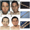
FIGURE 1
FIGURE 1
Clinical appearance of XP, XP/TTD, and TTD patients and polarizing microscopic examination of their hair. A–D: Clinical appearance of face and eyes. E–G: Microscopic examination of hair using polarized light [Liang et al., 2005]. A: Patient XP6BE, at age 18 years, had XP with neurological abnormalities [Robbins et al., 1974] and mutations in the XPD gene. Her face and neck show numerous freckle-like pigmented lesions of varying size and shape. There are scars from multiple surgical procedures. She had 25–50 basal cell and squamous cell carcinomas and two primary melanomas. The conjunctiva of her eyes show sunlight-induced inflammatory lesions. She died at the age of 29 years. B: Patient XPTTD268BE, age 18 years, has the XP/TTD complex and mutations in the XPD gene. He has the XP features of sunlight-induced freckle-like pigmented lesions of his face and neck and inflammatory lesions of the conjunctiva. His short, brittle hair and sparse eyebrows are features of TTD. E: Patient XPTTD268BE’s hair showed the TTD characteristic of alternating dark and light “tiger tail” bands with polarized light. C: Patient TTD351BE, age 9 years, has TTD and mutations in the XPD gene. He has short, brittle hair, sparse eyebrows, and ichthyosis of his upper chest. Note the absence of freckle-like pigmentation despite marked clinical sun sensitivity. F: Patient TTD351BE’s hair showed the TTD characteristic of alternating dark and light “tiger tail” bands with polarized light. D: Patient TTD331BE, age 27 years, has TTD and mutations in the TTDA gene. He had sun sensitivity without the pigmentation changes of XP.G: Patient TTD331BE’s hair showed the TTD characteristic of alternating dark and light “tiger tail” bands with polarized light.
FIGURE 2
FIGURE 2
TFIIH levels in unirradiated cells from XP, TTD, and XP/TTD patients measured by immunofluorescence and Western blotting. A: Normal cells (AG13145) were labeled with 0.8-μm beads (red arrows) and XP (XP-D) (Patients XP17BE and XP34BE), XP/ TTD (XP-D) (Patients XPTTD306BE and XPTTD260BE), and TTD (TTD-A) (Patient TTD331BE) and TTD (XP-D) (Patient TTD351BE) cells were labeled with 2-μm beads (yellow arrows) and co-cultured. Immunofluorescence staining was performed with antibodies to XPC, XPB, and XPD proteins. The intensity of staining of the patient cells is compared to that of the normal cells. XPC levels in all patient cells are comparable to normal cells. Levels of TFIIH subunits XPB and XPD are severely reduced in TTD cells of complementation groups XP-D and TTD-A, and reduced in XP/TTD cells of complementation group XP-D. B: Western blotting measurement of XPB protein levels in normal (AG13145); XP (XP-D) (Patients XP34BE and XP17BE); XP/TTD (XP-D) (Patient XPTTD306BE); and TTD (XP-D) (Patients TTD351BE and TTD355BE) cells. The ratio of the intensity of the XPB bands to that of the β-actin bands is indicted between the rows. C: Western blotting measurement of recombinant XPD-GFP protein expression (~110 kDa) in SV40-transformed TTD1VI-G3 [XPD-GFP]+ stably transfected fibroblasts compared to normal (GM0637) and TTD (XP-D) TTD1VI SV40-transformed cells. D: Western blot showing increased levels of XPD protein in clone G3 of SV40-transformed TTD1VI [XPD-GFP]+ stably transfected fibroblasts. Lanes were loaded with 50 or 100 μg of protein as indicated. The arrow indicates the migration of GFP-XPD. The lower bands are the size of endogenous XPD protein. The ratio of the intensity of the XPD bands to that of the β-actin bands is indicated between the rows.
FIGURE 3
FIGURE 3
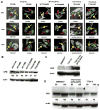
Recruitment of XPC and XPD proteins to localized DNA damage in XP, XP/TTD, and TTD cells at 0.1 hr, 0.5 hr, and 24 hr after UV irradiation. Normal cells (AG13145) were labeled with 0.8-μm latex beads (red arrows) and cells (from Patients XP17BE, XP6BE ER2-9, XPTTD306BE, TTD331BE, and TTD351BE) were labeled with 2-μm latex beads (yellow arrows). UV irradiation at 100 J/m2 was delivered through a 5-μm filter. Cells were fixed by 0.1 hr, 0.5 hr, or 24 hr postirradiation and immunostained with pairs of antibodies to simultaneously assess the location of DNA damage and NER proteins. The arrows indicate sites of localized immunostaining: normal (red arrows), patient (yellow arrows). Where necessary, images were captured at two exposure times to show any localization with low levels of protein (images separated by white line). Symbols below each image indicate localization (+) or nonlocalization (−) of NER proteins or photoproducts in normal cells/patient cells.
FIGURE 4
FIGURE 4
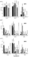
Frequency of XPC, XPD, and XPG NER protein localization in XP, XP/TTD, and TTD cells at 0.1 hr, 0.5 hr, and 24 hr after UV irradiation. A total of 50 nuclei were scored as being with (positive) or without (negative) DNA damage or NER protein localization at the indicated time points postirradiation. A: XPC localization. B: XPD localization. C: XPG localization. Where shown, error bars indicate the SEM of at least two independent counts.*p<0.001 significant difference between patient cell count and normal cell count at the equivalent time postirradiation. Bar numbers: 1, Patient AG13145; 2, Patient XP34BE; 3, Patient XP17BE; 4, Patient XPTTD306BE; 5, Patient TTD331BE; 6, Patient TTD351BE; 7, Patient TTD355BE; 8, Patient TTD1VI; and 9, Patient TTD1VI [XPD-GPF]+.
FIGURE 5
FIGURE 5
Recruitment of XPC, XPB, XPG, XPA, and XPF proteins to sites of localized UV damage in cells from Patient TTD6VI (XP-B) at 0.1hr, 0.5 hr, 3 hr, and 24 hr after UV irradiation. Normal AG13145 cells were labeled with 0.8-μm latex beads (red arrows) and cells from Patient TTD6VI (XP-B) were labeled with 2-μm latex beads (yellow arrows). Cells were fixed by 0.1hr, 0.5 hr, 3 hr, or 24 hr postirradiation and immunostained with pairs of antibodies to simultaneously assess the location of DNA damage and NER proteins. The arrows indicate sites of localized immunostaining: normal (red arrows), patient (yellow arrows). Symbols below each image indicate localization (+) or nonlocalization (−) of NER proteins or photoproducts in normal cells/patient cells. Experimental details are as in Fig.3.
FIGURE 6
FIGURE 6
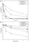
Repair of UV-induced 6-4PP and CPD in XP, TTD, and XP/TTD cells. Fibroblasts from normal (AG13146) or XP (XP-D) (Patient XP17BE); TTD (XP-D) (Patients TTD351BE and TTD355BE); and XP/TTD (XP-D) (Patient XPTTD306BE) cells were irradiated with 10 J/m2 UV and harvested at various times postirradiation. The percentage of the initial number of photoproducts was determined using an ELISA with antibodies specific to: (A) 6-4PP or (B) CPD. Error bars indicate the SEM of at least two independent experiments.
FIGURE 7
FIGURE 7
Schematic diagram of repair of UV-induced DNA damage (6-4PP and CPD) in normal, XP (XP-D or XP-B), and TTD (XP-D, XP-B, or TTD-A) cells. DNA is damaged by UV producing 6-4PP (yellow circles) and CPD (red circles). In normal cells (left column) by 0.1hr NER proteins are localized at the site of DNA damage. These proteins remain localized at 0.5 hr but by 24 hr the NER proteins are redistributed and the DNA damage has been excised in the normal cells. In contrast, in XP (XP-D or XP-B) cells (center column) by 0.1hr only XPC is visualized at the site of localized DNA damage. By 0.5 hr the NER proteins, including mutated XPD (dark pink oval with dashed border) and mutated XPB (light pink oval with dashed border), are recruited to the site of localized DNA damage. These proteins persist at 24 hr at sites of unrepaired 6-4PP and CPD. In TTD (XP-D, or XP-B or TTD-A) cells (right column) by 0.1hr only XPC is visualized at the site of localized DNA damage as in the XP cells. By 0.5 hr the NER proteins includingmutated XPD (dark pink oval with dotted border), mutated XPB (light pink oval with dotted border), and mutated TTDA (blue/green oval with dotted border) are recruited to the site of localized DNA damage in only a small proportion of the cells because of the instability of the TFIIH complex. At 24 hr the NER proteins have redistributed, CPD remain and 6-4PP are repaired. The NER proteins include DDB1-DDB2 (XPE) (purple ovals); XPC (light blue oval), which is complexed to HR23b and centrin 2; components of TFIIH: XPD (dark pink oval), XPB (light pink oval), TTDA (blue/green oval), and seven other proteins (light green ovals); and endonucleases XPF (dark gray oval) complexed to ERCC1 (light gray oval) and XPG (dark green oval). In the XP (XP-D or XP-B) cells the persistent NER proteins at the site of unrepaired 6-4PP and CPD may block progression of the translesion polymerase, pol eta, thereby creating a partial mimic of XP variant cells. This impaired pol eta function could result in increased mutagenesis of genes such as p53, which lead to cell cycle dysregulation that my eventually lead to cancer. In addition, mutations in TFIIH components have been shown to result in c-myc dysregulation with resulting cell-cycle dysregulation. The increased pigmentation in XP patients may be related to stimulation of tyrosinase activity via the UV-responsive transcription factor, Usf-1. In contrast, in TTD (XP-D, XP-D, or TTD-A) cells the unstable TFIIH complex is sufficient to repair 6-4PP but NER proteins do not persist at the site of unrepaired CPD. The translesion polymerase, pol eta can function normally to bypass the unrepaired DNA damage and thus does not induce an elevated frequency of cancer. (See discussion for more details.)

References

    1. Ahrens C, Grewe M, Berneburg M, Grether-Beck S, Quilliet X, Mezzina M, Sarasin A, Lehmann AR, Arlett CF, Krutmann J. Photocarcinogenesis and inhibition of intercellular adhesion molecule 1 expression in cells of DNA-repair-defective individuals. Proc Natl Acad Sci USA. 1997;94:6837–6841. - PMC - PubMed
    1. Andrews AD, Barrett SF, Robbins JH. Xeroderma pigmentosum neurological abnormalities correlate with colony-forming ability after ultraviolet radiation. Proc Natl Acad Sci USA. 1978;75:1984–1988. - PMC (V体育官网入口) - PubMed
    1. Barrett SF, Robbins JH, Tarone RE, Kraemer KH. Evidence for defective repair of cyclobutane pyrimidine dimers with normal repair of other DNA photoproducts in a transcriptionally active gene transfected into Cockayne syndrome cells. Mutat Res. 1991;255:281–291. - PubMed
    1. Berneburg M, Clingen PH, Harcourt SA, Lowe JE, Taylor EM, Green MH, Krutmann J, Arlett CF, Lehmann AR. The cancer-free phenotype in trichothiodystrophy is unrelated to its repair defect. Cancer Res. 2000;60:431–438. - PubMed
    1. Bootsma D, Hoeijmakers JH. DNA repair. Engagement with transcription. Nature. 1993;363:114–115. - PubMed

MeSH terms