"VSports在线直播" Skip to main page content
U.S. flag

An official website of the United States government

Dot gov

The VSports app下载. gov means it’s official. Federal government websites often end in . gov or . mil. Before sharing sensitive information, make sure you’re on a federal government site. .

Https

The site is secure. The https:// ensures that you are connecting to the official website and that any information you provide is encrypted and transmitted securely. V体育官网.

. 2008 May 1;68(9):3429-39.
doi: 10.1158/0008-5472.CAN-07-5867.

VSports app下载 - Akt3 and mutant V600E B-Raf cooperate to promote early melanoma development

Affiliations

Akt3 and mutant V600E B-Raf cooperate to promote early melanoma development

Mitchell Cheung et al. Cancer Res. .

Abstract

B-Raf is the most mutated gene in melanoma; however, the mechanism through which it promotes early melanomas remains uncertain. Most nevi contain activated (V600E)B-Raf but few develop into melanoma, and expression in melanocytes is inhibitory with low protein levels present in surviving cells, suggesting unknown cooperative oncogenic events are necessary for melanoma development VSports手机版. Because many melanomas have (V600E)B-Raf and active Akt3, it is possible that these proteins cooperatively facilitate melanocyte transformation. In this study, Akt3 is shown to phosphorylate (V600E)B-Raf to lower its activity as well as that of the downstream mitogen-activated protein kinase (MAPK) pathway to levels promoting early melanoma development. Expression of active Akt3 in early melanoma cells containing (V600E)B-Raf reduced MAPK signaling and promoted anchorage-independent growth. Furthermore, expression of both (V600E)B-Raf and active Akt3 in melanocytes promoted a transformed phenotype. Mechanistically, aberrant Akt3 activity in early melanomas serves to phosphorylate Ser(364) and Ser(428) on (V600E)B-Raf to reduce activity of (V600E)B-Raf to levels that promote rather than inhibit proliferation, which aids melanocytic transformation. Inhibition of (V600E)B-Raf or Akt3 in advanced melanoma cells in which both pathways were active reduced anchorage-independent growth and tumor development in a cooperatively acting manner. Inhibition of Akt3 alone in these cells led to increased MAPK signaling. In summary, these results suggest that activating B-Raf mutations initially promote nevi development, but the resulting high, intense activation of the MAPK pathway inhibits further tumor progression requiring Akt3 activation to bypass this barrier and aid melanoma development. .


Figures

Figure 1
Figure 1
Crosstalk between the Akt3 and MAP Kinase pathway occurs in melanoma cells but not normal melanocytes. Phosphorylated Akt and Erk levels of melanocytes (A.) and UACC 903 (B.) treated with or without LY-294002. Graphs represent densitometric scans of pAkt and pErk Western blots normalized to a α-enolase loading control. Cells were starved 24 hours, treated with or without LY-294002 for 20 minutes, then serum stimulated before harvesting lysates at different time points for Western blot analysis. Analysis shows LY-294002 abrogates activation of Akt (pAkt) and consequently increase Erk activation (phosphorylation) in melanoma cells but not in human melanocytes. C, Inhibition of Akt activation results in higher pErk levels in melanoma cell lines but not in melanocytes. Western blots are shown for pAkt and pErk levels in normal human melanocytes and two human metastatic melanoma cell lines, UACC 903 and SK-MEL-24. Cells were starved 24 hours, then untreated or treated with DMSO or 50 mM LY-294002 for 20 minutes, and then stimulated with growth factors or serum in media and protein lysates harvested for Western blot analysis. The two metastatic melanoma cell lines showed evidence that LY-294002 treatment increased pErk levels, which did not occur in normal human melanocytes. D, Inhibition of Akt3 increased pErk levels in melanoma cells. Normal melanocytes were transfected with scrambled siRNA, or siRNA targeting PTEN or Akt3 alone or in combination. After 48 h, cell lysates were analyzed for PTEN, Akt3, pAkt and pErk levels. Inhibiting Akt3 protein expression has no effect on pErk levels. UACC 903 cells were transfected with buffer, scrambled siRNA, or Akt3 targeting siRNA, starved 24 hours, then stimulated with media containing serum for 30 or 60 minutes and protein lysates harvested for Western blot analysis. siRNA-mediated inhibition of Akt3 increased pErk levels compared to buffer or scrambled siRNA controls. α-enolase served as a control for protein loading.
Figure 1
Figure 1
Crosstalk between the Akt3 and MAP Kinase pathway occurs in melanoma cells but not normal melanocytes. Phosphorylated Akt and Erk levels of melanocytes (A.) and UACC 903 (B.) treated with or without LY-294002. Graphs represent densitometric scans of pAkt and pErk Western blots normalized to a α-enolase loading control. Cells were starved 24 hours, treated with or without LY-294002 for 20 minutes, then serum stimulated before harvesting lysates at different time points for Western blot analysis. Analysis shows LY-294002 abrogates activation of Akt (pAkt) and consequently increase Erk activation (phosphorylation) in melanoma cells but not in human melanocytes. C, Inhibition of Akt activation results in higher pErk levels in melanoma cell lines but not in melanocytes. Western blots are shown for pAkt and pErk levels in normal human melanocytes and two human metastatic melanoma cell lines, UACC 903 and SK-MEL-24. Cells were starved 24 hours, then untreated or treated with DMSO or 50 mM LY-294002 for 20 minutes, and then stimulated with growth factors or serum in media and protein lysates harvested for Western blot analysis. The two metastatic melanoma cell lines showed evidence that LY-294002 treatment increased pErk levels, which did not occur in normal human melanocytes. D, Inhibition of Akt3 increased pErk levels in melanoma cells. Normal melanocytes were transfected with scrambled siRNA, or siRNA targeting PTEN or Akt3 alone or in combination. After 48 h, cell lysates were analyzed for PTEN, Akt3, pAkt and pErk levels. Inhibiting Akt3 protein expression has no effect on pErk levels. UACC 903 cells were transfected with buffer, scrambled siRNA, or Akt3 targeting siRNA, starved 24 hours, then stimulated with media containing serum for 30 or 60 minutes and protein lysates harvested for Western blot analysis. siRNA-mediated inhibition of Akt3 increased pErk levels compared to buffer or scrambled siRNA controls. α-enolase served as a control for protein loading.
Figure 2
Figure 2
V600EB-Raf and Akt3 regulate anchorage independent growth of early melanoma cells. A, WM35 has constitutive activation of MAP kinase pathway signaling due to presence of V600EB-Raf but normal Akt activity. Densitometric quantitation of pAkt and pErk levels from Western blots of the WM35 cell line following 24 hours serum starvation followed by stimulation, shows constitutively high levels of pErk despite serum starvation, which is caused by V600EB-Raf activating the MAP kinase signaling cascade. In contrast, the Akt activation profile is similar to that observed for melanocytes. B, Inhibition of pAkt in WM35 cells using LY-294002 resulted in higher pErk while expression of active Akt3 decreased pErk levels. Treatment with LY-294002 led to a reduction in pAkt and a corresponding increase in pErk levels. Ectopic expression of active Akt3 decreased pErk levels. WM35 cells were nucleofected with Akt3 expressing constructs and following two days recovery, cells were starved 24 hours, and then stimulated with serum containing media and protein lysates collected for Western blot analysis. Ectopic myristoylated-Akt3 expression led to decreased pErk levels. C, Inhibition of B-Raf decreases anchorage independent growth of early melanoma cells. WM35 cells were nucleofected with siRNA targeting V600EB-Raf (100 and 200 pmoles), scrambled siRNA, or a water control. Cells were then plated onto PolyHEMA coated 96 well plates to measure effect on anchorage independent growth. MTS assay, 3 days later, was used to quantify viable cell number. Western blot showed efficient knockdown of B-Raf protein expression and decreased pErk levels. α-enolase served as a control for protein loading. D, Ectopic expression of active Akt3 enhanced anchorage independent growth. Viability of WM35 cells nucleofected with Akt3 is shown along with Western blot analysis of cell lysates. WM35 cells were nucleofected with various Akt3 expressing constructs: empty vector, WT-Akt3, Dead-Akt3 and active Akt3: myristoylated-Akt3 (myr-Akt3) or E40K-Akt3 alone or in combination with a siRNA targeting PRAS40 protein. After nucleofection, cells were plated directly onto PolyHEMA coated 96 well plates and following 3 days of growth in selection media, an MTS assay was used to quantify cell viability. Myr-Akt3 significantly increased cell growth compared to less active E40K-Akt3 that had a more modest effect. Changes in cell growth corresponded directly to pAkt level, which is shown by Western blot analysis and quantified by densitometry. Knocking down PRAS40 had no significant effect on cell viability. HA probed Western blot shows equal ectopic expression of the different Akt3 proteins.
Figure 2
Figure 2
V600EB-Raf and Akt3 regulate anchorage independent growth of early melanoma cells. A, WM35 has constitutive activation of MAP kinase pathway signaling due to presence of V600EB-Raf but normal Akt activity. Densitometric quantitation of pAkt and pErk levels from Western blots of the WM35 cell line following 24 hours serum starvation followed by stimulation, shows constitutively high levels of pErk despite serum starvation, which is caused by V600EB-Raf activating the MAP kinase signaling cascade. In contrast, the Akt activation profile is similar to that observed for melanocytes. B, Inhibition of pAkt in WM35 cells using LY-294002 resulted in higher pErk while expression of active Akt3 decreased pErk levels. Treatment with LY-294002 led to a reduction in pAkt and a corresponding increase in pErk levels. Ectopic expression of active Akt3 decreased pErk levels. WM35 cells were nucleofected with Akt3 expressing constructs and following two days recovery, cells were starved 24 hours, and then stimulated with serum containing media and protein lysates collected for Western blot analysis. Ectopic myristoylated-Akt3 expression led to decreased pErk levels. C, Inhibition of B-Raf decreases anchorage independent growth of early melanoma cells. WM35 cells were nucleofected with siRNA targeting V600EB-Raf (100 and 200 pmoles), scrambled siRNA, or a water control. Cells were then plated onto PolyHEMA coated 96 well plates to measure effect on anchorage independent growth. MTS assay, 3 days later, was used to quantify viable cell number. Western blot showed efficient knockdown of B-Raf protein expression and decreased pErk levels. α-enolase served as a control for protein loading. D, Ectopic expression of active Akt3 enhanced anchorage independent growth. Viability of WM35 cells nucleofected with Akt3 is shown along with Western blot analysis of cell lysates. WM35 cells were nucleofected with various Akt3 expressing constructs: empty vector, WT-Akt3, Dead-Akt3 and active Akt3: myristoylated-Akt3 (myr-Akt3) or E40K-Akt3 alone or in combination with a siRNA targeting PRAS40 protein. After nucleofection, cells were plated directly onto PolyHEMA coated 96 well plates and following 3 days of growth in selection media, an MTS assay was used to quantify cell viability. Myr-Akt3 significantly increased cell growth compared to less active E40K-Akt3 that had a more modest effect. Changes in cell growth corresponded directly to pAkt level, which is shown by Western blot analysis and quantified by densitometry. Knocking down PRAS40 had no significant effect on cell viability. HA probed Western blot shows equal ectopic expression of the different Akt3 proteins.
Figure 3
Figure 3
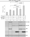
V600EB-Raf and Akt3 cooperate in melanocyte transformation. A, Melan-a transformation, following ectopic V600EB-Raf and Akt3 expression, was quantified by determined effect on anchorage independent growth. Melan-a cells were nucleofected with various combinations of V600EB-Raf and Akt3 expression plasmids (resistant to blasticidin and puromycin, respectively) and then directly plated onto PolyHEMA coated 96 well plates. Transfected cells were selected for blasticidin and puromycin resistance for three days followed by MTS assay to quantify cell viability. Results of a representative MTS assay are shown indicating effects of V600EB-Raf and Akt3 expression on anchorage independent growth. Nucleofections of either V600EB-Raf or myr-Akt3 led to increased cell viability; however, co-expression of both genes resulted in a more statistically significant effect. Western blots indicate changes in signaling pathways following ectopic expression of B-Raf and Akt3. Error bars; mean ± S.D. B, In vitro B-Raf kinase assay showing activity of V600EB-Raf compared to protein in which phosphorylation sites are converted to alanine S364A and S428A. Upper blot show autoradiography film of 32P labeled Mek substrate while lower shows a Western blot quantifying the amount of immunoprecipitated B-Raf protein used for the kinase assay. The graph represents average densitometry analysis (±S.D.) of three independent experiments, where values indicate level of labeled Mek normalized to the amount of HA B-Raf, relative to Dead B-Raf set as the background control. C, Akt3 phosphorylates V600EB-Raf on serines 364 and 428. Quantitation of B-Raf phosphorylation using densitometric scans of Western blot that were normalized to total immunoprecipitated HA B-Raf. Both wild type and V600EB-Raf were phosphorylated by Akt3. Conversion of serine 364 to alanine reduced levels of V600EB-Raf phosphorylation, while mutation of both serines 364 and 428 completely abrogated phosphorylation by Akt3. Graph is the average of three independent experiments; error bars, mean ±S.D.
Figure 3
Figure 3
V600EB-Raf and Akt3 cooperate in melanocyte transformation. A, Melan-a transformation, following ectopic V600EB-Raf and Akt3 expression, was quantified by determined effect on anchorage independent growth. Melan-a cells were nucleofected with various combinations of V600EB-Raf and Akt3 expression plasmids (resistant to blasticidin and puromycin, respectively) and then directly plated onto PolyHEMA coated 96 well plates. Transfected cells were selected for blasticidin and puromycin resistance for three days followed by MTS assay to quantify cell viability. Results of a representative MTS assay are shown indicating effects of V600EB-Raf and Akt3 expression on anchorage independent growth. Nucleofections of either V600EB-Raf or myr-Akt3 led to increased cell viability; however, co-expression of both genes resulted in a more statistically significant effect. Western blots indicate changes in signaling pathways following ectopic expression of B-Raf and Akt3. Error bars; mean ± S.D. B, In vitro B-Raf kinase assay showing activity of V600EB-Raf compared to protein in which phosphorylation sites are converted to alanine S364A and S428A. Upper blot show autoradiography film of 32P labeled Mek substrate while lower shows a Western blot quantifying the amount of immunoprecipitated B-Raf protein used for the kinase assay. The graph represents average densitometry analysis (±S.D.) of three independent experiments, where values indicate level of labeled Mek normalized to the amount of HA B-Raf, relative to Dead B-Raf set as the background control. C, Akt3 phosphorylates V600EB-Raf on serines 364 and 428. Quantitation of B-Raf phosphorylation using densitometric scans of Western blot that were normalized to total immunoprecipitated HA B-Raf. Both wild type and V600EB-Raf were phosphorylated by Akt3. Conversion of serine 364 to alanine reduced levels of V600EB-Raf phosphorylation, while mutation of both serines 364 and 428 completely abrogated phosphorylation by Akt3. Graph is the average of three independent experiments; error bars, mean ±S.D.
Figure 4
Figure 4
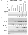
V600EB-Raf and Akt3 mediated transformation requires Akt3 phosphorylation of V600EB-Raf. A, Representative graph showing anchorage independent growth mediated by V600EB-Raf and Akt3 coexpression. Melan-a cells were nucleofected with different combinations of S364A/S428A V600EB-Raf (AA-V600EB-Raf) or V600EB-Raf and Akt3 expressing plasmids, and plated onto PolyHEMA coated 96 well plates. Transfected cells were then selected for three days for purimycin resistance followed by MTS assay to measure cell viability. Conversion of the two serine phosphorylation sites on V600EB-Raf to alanines (S364A/S428A) led to significantly decreased anchorage independent growth mediated by V600EB-Raf and Akt3 expression; error bars, mean ±S.D. of four independent experiments. B, Western blots showing changes in signaling pathways following ectopic expression of B-Raf and Akt3. Increased cyclin D1 levels are associated with coexpression of V600EB-Raf and Akt3.
Figure 5
Figure 5
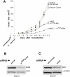
Targeting V600EB-Raf and Akt3 dramatically retards melanoma tumorigenesis. A, Nucleofection of siRNA against both B-Raf and Akt3 into UACC 903 cells led to a statistically significant reduction of tumor development in animals. The cells were nucleofected with siRNA, allowed to recover in culture for 1.5 days, and then injected subcutaneously into nude mice. Tumor size was measured on alternate days up to day 17.5. Inhibition of Akt3 or B-Raf alone led to a significant reduction in growth compared to controls; however, targeting both proteins led to an even more dramatic reduction compared to targeting Akt3 or B-Raf individually. Bars represent mean ± S.E. B, Western blot of UACC 903 tumors isolated from mice 8 days after nucleofection with siRNA targeting V600EB-Raf. siRNA against B-Raf significantly reduced level of B-Raf protein compared to control cells nucleofected with siRNA to C-Raf. C, Western blot of UACC 903 tumors isolated from mice 8 days after nucleofection with siRNA targeting Akt3. Akt3 protein level was significantly reduced with siRNA targeting Akt3 compared to cells nucleofected with a scrambled siRNA control.
Figure 6
Figure 6
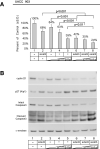
Simultaneously targeting V600EB-Raf and Akt3 inhibits in vitro melanoma growth in an additively cooperating manner. A, In vitro anchorage independent growth of UACC 903 cells nucleofected with siRNA targeting V600EB-Raf and Akt3 in cell culture media containing 0% serum. UACC 903 cells nucleofected with siRNA were plated onto PolyHEMA coated 96 well plates, allowed to grow for 3 days in DMEM containing no serum and cell viability quantified by MTS assay. UACC 903 cells nucleofected with no siRNA (lane 1) was set as 100% and others compared to this value. Targeting both B-Raf and Akt3 resulted in statistically significant inhibition of growth compared to cells in which B-Raf or Akt3 had been targeted individually. B, Western blots showing effects of targeting V600EB-Raf and Akt3 on cyclin D1, cleaved caspase-3 and p27Kip1 protein levels. siRNA-mediated inhibition of V600EB-Raf and Akt3 led to significant reductions in cyclin D1 and corresponding increases in cleaved caspase3 and p27Kip1 protein levels compared to cells in which proteins had been targeted individually.

References

    1. Cancer Facts and Figures. American Cancer Society; Atlanta: 2005.
    1. Smalley KS, Herlyn M. Targeting intracellular signaling pathways as a novel strategy in melanoma therapeutics. Ann N Y Acad Sci. 2005;1059:16–25. - PubMed (VSports app下载)
    1. Hsu MY, Meier F, Herlyn M. Melanoma development and progression: a conspiracy between tumor and host. Differentiation. 2002;70:522–36. - PubMed
    1. Chudnovsky Y, Khavari PA, Adams AE. Melanoma genetics and the development of rational therapeutics. J Clin Invest. 2005;115:813–24. - PMC - PubMed
    1. Kantrow SM, Boyd AS, Ellis DL, et al. Expression of activated Akt in benign nevi, Spitz nevi and melanomas. J Cutan Pathol. 2007;34:593–6. - PMC - PubMed

Publication types

MeSH terms