Skip to main page content
U.S. flag

An official website of the United States government

Dot gov

The . gov means it’s official VSports app下载. Federal government websites often end in . gov or . mil. Before sharing sensitive information, make sure you’re on a federal government site. .

Https

The site is secure V体育官网. The https:// ensures that you are connecting to the official website and that any information you provide is encrypted and transmitted securely. .

. 2008 Jul;28(13):4215-26.
doi: 10.1128/MCB.00867-07. Epub 2008 Apr 21.

A Rictor-Myo1c complex participates in dynamic cortical actin events in 3T3-L1 adipocytes

Affiliations

A Rictor-Myo1c complex participates in dynamic cortical actin events in 3T3-L1 adipocytes

V体育ios版 - G Nana Hagan et al. Mol Cell Biol. 2008 Jul.

Abstract

Insulin signaling through phosphatidylinositol 3-kinase (PI 3-kinase) activates the protein kinase Akt through phosphorylation of its threonine 308 and serine 473 residues by the PDK1 protein kinase and the Rictor-mammalian target of rapamycin complex (mTORC2), respectively. Remarkably, we show here that the Rictor protein is also present in cultured adipocytes in complexes containing Myo1c, a molecular motor that promotes cortical actin remodeling. Interestingly, the Rictor-Myo1c complex is biochemically distinct from the previously reported mTORC2 and can be immunoprecipitated independently of mTORC2. Furthermore, while RNA interference-directed silencing of Rictor results in the expected attenuation of Akt phosphorylation at serine 473, depletion of Myo1c is without effect. In contrast, loss of either Rictor or Myo1c inhibits phosphorylation of the actin filament regulatory protein paxillin at tyrosine 118 VSports手机版. Furthermore, Myo1c-induced membrane ruffling of 3T3-L1 adipocytes is also compromised following Rictor knockdown. Interestingly, neither the mTORC2 inhibitor rapamycin nor the PI 3-kinase inhibitor wortmannin affects paxillin tyrosine 118 phosphorylation. Taken together, our findings suggest that the Rictor-Myo1c complex is distinct from mTORC2 and that Myo1c, in conjunction with Rictor, participates in cortical actin remodeling events. .


Figures (VSports在线直播)

FIG. 1.
FIG. 1.
Rictor coimmunoprecipitates with the unconventional myosin Myo1c. (A) Identification of Myo1c as an immunoprecipitating partner of Rictor. Lysates from WT and Rictor-deficient MEFs (from Rictor knockout mice) were subjected to control rabbit IgG or Rictor polyclonal antibody IP. After electrophoresis of samples in SDS-polyacrylamide gels, the gels were silver stained. The bands in the regions shown with solid triangles were excised and identified by mass spectrometry analysis. The proteins that immunoprecipitated with Rictor from WT MEFs but were absent in samples treated with control rabbit IgG and in samples derived from Rictor knockout MEFs were analyzed further. As shown in the top panel, the upper two bands were mTOR (above MW of 212,000) and Rictor (below MW of 212,000). MWs in figures are thousands. (B) The boxed region of the gel in panel A was enhanced for better visualization of a band in the region around an MW of 116,000. This band was identified as Myo1c. (C) Quantification of results. Whereas five Myo1c peptides were found following IP of Rictor from WT MEFs, no Myo1c peptides were found following IP of Rictor from Rictor-deficient MEFs. Other proteins were identified, and the numbers of peptides found following Rictor IP from WT and Rictor knockout (KO) MEFs are shown. (D) IP between endogenous Myo1c and Rictor in 3T3-L1 adipocytes subjected to 0.3% CHAPS lysis conditions. Differentiated 3T3-L1 adipocytes were serum starved or stimulated with 100 nM insulin for 15 min and then lysed with 0.3% CHAPS lysis buffer. IP studies were then performed using the 0.3% CHAPS 3T3-L1 adipocyte lysates and antibodies to mTOR, Rictor, Myo1c, and nonimmune rabbit IgG (pull down). Membranes were then blotted for mTOR, Rictor, Myo1c, and Raptor. (E) IP of endogenous Myo1c and Rictor in 3T3-L1 adipocytes under 1% Triton X-100 lysis conditions. Differentiated 3T3-L1 adipocytes were serum starved or stimulated with 100 nM insulin for 15 min and then lysed with 1% Triton X-100 lysis buffer. IP studies were then performed using the 1% Triton X-100 3T3-L1 adipocyte lysates and antibodies to mTOR, Rictor, Myo1c, and nonimmune rabbit IgG (pull down). Membranes were then blotted for mTOR, Rictor, Myo1c, and Raptor. The results shown are representative of three independent experiments. In all 3T3-L1 adipocyte IP experiments 50 μg of total protein was used for lysate blots and 2 mg of total protein was used for IP experiments. −, basal conditions; +, 100 nM insulin stimulation.
FIG. 2.
FIG. 2.
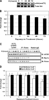
Rictor-Myo1c complex is insensitive to chronic rapamycin treatment. (A) Effect of prolonged rapamycin treatment on Akt phosphorylation (serine 473) in 3T3-L1 adipocytes. Differentiated 3T3-L1 adipocytes were treated with 100 nM rapamycin for 0, 1, 24, 48, or 72 h. Adipocytes from each time point were lysed with 0.3% CHAPS lysis buffer, and lysates were immunoblotted for Akt phosphorylation at serine 473 as well as for total Akt. The images shown are representative blots from three independent experiments. (B) Quantitative assessment of the results in panel A, based on densitometry analysis of three independent experiments. (C) Effect of prolonged rapamycin treatment on Rictor-mTOR and Rictor-Myo1c complex stability. IP studies were performed by immunoprecipitating Rictor and immunoblotting for mTOR or Myo1c, using the 0.3% CHAPS 3T3-L1 adipocyte lysates from panel A. The images shown are representative blots from three independent experiments. (D) Quantitative assessment of the results in panel C, based on densitometry analysis of three independent experiments. In all 3T3-L1 adipocyte IP experiments, 50 μg of total protein was used for lysate blots and 2 mg of total protein was used for IP experiments. R, rapamycin treatment; pAkt, phospho-Akt (serine 473); IB, immunoblot. *, P < 0.05.
FIG. 3.
FIG. 3.
Rictor coimmunoprecipitates with the motor domain of Myo1c. (A) Triple-HA-tagged Myo1c constructs are shown. In addition to the full-length Myo1c(motor+IQ+tail) construct, Myo1c(motor+IQ), Myo1c(IQ+tail), and Myo1c(tail) constructs were prepared for cotransfection and IP experiments with a myc-tagged Rictor construct. (B) IP studies of myc-Rictor and 3×HA-Myo1c constructs. HEK 293T cells were cotransfected with myc-Rictor and either 3×HA-Myo1c(full), 3×HA-Myo1c(motor+IQ), 3×HA-Myo1c(IQ+tail), or 3×HA-Myo1c (tail). At 48 h posttransfection, cells were lysed with 1% Triton X-100 lysis buffer. Lysates were immunoprecipitated with the myc tag and immunoblotted for the HA or myc tag. Nonimmune rabbit IgG was used as a control for IP. The image shown is a representative blot from three independent experiments. (C) Assessment of latrunculin B effect on cortical actin integrity. 3T3-L1 adipocytes were incubated with or without 2 μM latrunculin B. At 3 h postincubation, cells were fixed with 4% paraformaldehyde, permeabilized with 0.1% Triton X-100, and stained with rhodamine-phalloidin for cortical actin analysis. The images shown are representative frames of cells for each condition. Bars, 10 μm. (D) IP studies of endogenous Rictor and Myo1c following latrunculin B treatment. 3T3-L1 adipocytes were incubated without (−) or with (+) 2 μM latrunculin B. At 3 h postincubation, cells were lysed with 0.1% Triton X-100 lysis buffer. Lysates were immunoprecipitated with Rictor and immunoblotted for Myo1c. Nonimmune rabbit IgG was used as a control for IP. The images shown are representative blots from three independent experiments. MWs in the figure are thousands.
FIG. 4.
FIG. 4.
Rictor depletion but not Myo1c depletion results in decreased Akt (serine 473) phosphorylation. (A) siRNA-mediated Rictor and Myo1c silencing. Differentiated 3T3-L1 adipocytes were transfected with either scrambled siRNAs (S), siRNAs to Rictor (R), or siRNAs to Myo1c (M). At 72 h posttransfection, the adipocytes were serum starved for 3 h and then left serum starved (0.0 nM insulin) or stimulated with 0.1 nM, 1 nM, 10 nM, or 100 nM insulin for 15 min. Lysates from each condition were analyzed by immunoblotting to determine the levels of Rictor depletion, Myo1c depletion, Akt phosphorylation (serine 473), and total Akt. The images shown are representative immunoblots from three independent experiments. (B) Quantitative assessment of the results in panel A, based on densitometry analysis of three independent experiments. Significance levels for comparison of Myo1c siRNA-transfected cells to Rictor siRNA-transfected cells are as follows: *, P < 0.05; **, P < 0.01. Significance levels for comparison of scrambled siRNA-transfected cells to Rictor siRNA-transfected cells are as follows: #, P < 0.05; ##, P < 0.01.
FIG. 5.
FIG. 5.
Rictor and Myo1c both regulate paxillin phosphorylation at tyrosine 118, in an mTOR- and PI3K-independent manner. (A) Rictor and Myo1c silencing compromises paxillin phosphorylation at tyrosine 118 (Y118). Differentiated 3T3-L1 adipocytes were transfected with either scrambled siRNAs, siRNAs to Rictor, or siRNAs to Myo1c. At 72 h posttransfection, the adipocytes were lysed in 1% SDS buffer and immunoblots were performed to assess Rictor, Myo1c, phospho-paxillin (Y118), and total paxillin levels. The images shown are representative blots from three independent experiments. (B) Effect of prolonged rapamycin treatment on phospho-paxillin level. Differentiated 3T3-L1 adipocytes were either left unstimulated and untreated (−/−), left unstimulated but treated with 100 nM rapamycin for 72 h (−/+), stimulated with 100 nM insulin for 15 min but left untreated (+/−), or stimulated with 100 nM insulin for 15 min and treated with 100 nM rapamycin for 72 h (+/+). In all cases where it was present, insulin was added for the final 15 min of the experiment. Following stimulation and treatment, the adipocytes were lysed in 1% SDS and immunoblots were performed to assess phospho-paxillin (Y118), total paxillin, phospho-Akt (S473), and total Akt levels. The images shown are representative blots from three independent experiments. (C) Effect of wortmannin treatment on phospho-paxillin level. Differentiated 3T3-L1 adipocytes were either left unstimulated and untreated (−/−), treated with 100 nM wortmannin for 30 min but left unstimulated (−/+), left untreated but stimulated with 100 nM insulin for 15 min (+/−), or treated with 100 nM wortmannin for 30 min and stimulated with 100 nM insulin for 15 min (+/+). Following treatment and stimulation, the adipocytes were lysed and immunoblots were performed to assess phospho-paxillin (Y118), total paxillin, phospho-Akt (S473), and total Akt levels. The images shown are representative blots from three independent experiments. Ins, 100 nM insulin; Rap, 100 nM rapamycin; Wort, 100 nM wortmannin.
FIG. 6.
FIG. 6.
Rictor depletion attenuates Myo1c-induced, insulin-independent membrane ruffling in 3T3-L1 adipocytes. (A) Rictor knockdown in 3T3-L1 adipocytes cotransfected with GFP-Myo1c and scrambled siRNA or Rictor siRNA. Differentiated 3T3-L1 adipocytes were cotransfected with GFP empty vector (GFP-E/V) or GFP-Myo1c and scrambled siRNA or Rictor siRNA. At 72 h posttransfection, a subset of these cells were lysed in 1% SDS and immunoblots were performed to assess Rictor and Akt (loading control) levels. (B) Rictor depletion abrogates Myo1c-induced, insulin-independent membrane ruffling in 3T3-L1 adipocytes (live-cell analysis). Differentiated 3T3-L1 adipocytes were cotransfected with GFP empty vector or GFP-Myo1c and scrambled siRNA or Rictor siRNA. At 72 h posttransfection, a subset of these cells were serum starved for 5 h and then incubated in Krebs-Ringer-HEPES buffer containing 0.5% bovine serum albumin and 1 mM sodium pyruvate. Membrane ruffling was observed by live monitoring of the cells for 10 min, with imaging done at 5-s intervals. The images shown are bright-field images and images at three time frames, each 5 min apart, for a representative GFP empty vector- and scrambled siRNA-transfected adipocyte, a GFP-Myo1c- and scrambled siRNA-transfected adipocyte, a GFP empty vector- and Rictor siRNA-transfected adipocyte, and a GFP-Myo1c- and Rictor siRNA-transfected adipocyte. Scr, scrambled siRNA; Ric, Rictor siRNA. Bars, 10 μm.
FIG. 7.
FIG. 7.
Rictor depletion abrogates Myo1c-induced, insulin-independent membrane ruffling in 3T3-L1 adipocytes (fixed-cell analysis). (A) Differentiated 3T3-L1 adipocytes were cotransfected with GFP empty vector or GFP-Myo1c and scrambled siRNA or Rictor siRNA and then seeded on glass coverslips. At 72 h posttransfection, these cells were serum starved for 5 h, fixed with 4% paraformaldehyde, permeabilized with 0.1% Triton X-100, and stained with rhodamine-phalloidin for membrane ruffle analysis. The images show representative adipocytes for each condition. Bars, 10 μm. (B) For each condition, the percentage of ruffled cells was determined by scoring the number of GFP-Myo1c-positive cells that had membrane ruffles against the total number of cells counted. At least 60 cells were evaluated in each experiment. The averaged data from three independent experiments are shown, with standard deviations. **, P < 0.01. (C) Effect of Rictor knockdown on Myo1c-induced, insulin-independent membrane ruffle size. 3T3-L1 adipocytes were transfected and prepared as described above. For each condition, membrane ruffle size was determined by measuring the size of discernible membrane ruffles for GFP-Myo1c-positive cells under each condition and dividing it by the total size of the cell, as determined using Axiovision software. For each cell evaluated, the GFP intensity was determined and the average ruffle size/total cell size ratio from each condition was normalized to the average GFP intensity of all evaluated cells under each condition. At least 60 cells were assessed in each experiment. The box-and-whisker plots show (from the top down) maximum, third quartile, median, first quartile, and minimum values. **, P < 0.01.
FIG. 7.
FIG. 7.
Rictor depletion abrogates Myo1c-induced, insulin-independent membrane ruffling in 3T3-L1 adipocytes (fixed-cell analysis). (A) Differentiated 3T3-L1 adipocytes were cotransfected with GFP empty vector or GFP-Myo1c and scrambled siRNA or Rictor siRNA and then seeded on glass coverslips. At 72 h posttransfection, these cells were serum starved for 5 h, fixed with 4% paraformaldehyde, permeabilized with 0.1% Triton X-100, and stained with rhodamine-phalloidin for membrane ruffle analysis. The images show representative adipocytes for each condition. Bars, 10 μm. (B) For each condition, the percentage of ruffled cells was determined by scoring the number of GFP-Myo1c-positive cells that had membrane ruffles against the total number of cells counted. At least 60 cells were evaluated in each experiment. The averaged data from three independent experiments are shown, with standard deviations. **, P < 0.01. (C) Effect of Rictor knockdown on Myo1c-induced, insulin-independent membrane ruffle size. 3T3-L1 adipocytes were transfected and prepared as described above. For each condition, membrane ruffle size was determined by measuring the size of discernible membrane ruffles for GFP-Myo1c-positive cells under each condition and dividing it by the total size of the cell, as determined using Axiovision software. For each cell evaluated, the GFP intensity was determined and the average ruffle size/total cell size ratio from each condition was normalized to the average GFP intensity of all evaluated cells under each condition. At least 60 cells were assessed in each experiment. The box-and-whisker plots show (from the top down) maximum, third quartile, median, first quartile, and minimum values. **, P < 0.01.
FIG. 8.
FIG. 8.
Working model suggesting that Rictor may function in two pathways. Our data suggest that Myo1c and Rictor form a complex in 3T3-L1 adipocytes that is biochemically and functionally distinct from the Rictor-mTOR complex. While the Rictor-mTOR complex (I) that phosphorylates and activates Akt is rapamycin sensitive, our data suggest that the Rictor-Myo1c complex (II) acts in an insulin-, rapamycin-, and wortmannin-insensitive manner to participate in dynamic cortical actin events.

References

    1. Avruch, J., K. Hara, Y. Lin, M. Liu, X. Long, S. Ortiz-Vega, and K. Yonezawa. 2006. Insulin and amino-acid regulation of mTOR signaling and kinase activity through the Rheb GTPase. Oncogene 256361-6372. - V体育官网 - PubMed
    1. Bar-Sagi, D., and A. Hall. 2000. Ras and Rho GTPases: a family reunion. Cell 103227-238. - PubMed
    1. Bose, A., A. Guilherme, S. I. Robida, S. M. C. Nicoloro, Q. L. Zhou, Z. Y. Jiang, D. P. Pomerleau, and M. P. Czech. 2002. Glucose transporter recycling in response to insulin is facilitated by myosin Myo1c. Nature 420821-824. - PubMed
    1. Bose, A., S. Robida, P. S. Furcinitti, A. Chawla, K. Fogarty, S. Corvera, and M. P. Czech. 2004. Unconventional myosin Myo1c promotes membrane fusion in a regulated exocytic pathway. Mol. Cell. Biol. 245447-5458. - PMC - PubMed
    1. Reference deleted.

Publication types

MeSH terms

Substances