<b lang="zTSgCyJ"></b> Skip to main page content
U.S. flag

An official website of the United States government

Dot gov

The VSports app下载. gov means it’s official. Federal government websites often end in . gov or . mil. Before sharing sensitive information, make sure you’re on a federal government site. .

Https

The site is secure. The https:// ensures that you are connecting to the official website and that any information you provide is encrypted and transmitted securely. V体育官网.

. 2008 Apr;14(4):421-8.
doi: 10.1038/nm1743. Epub 2008 Mar 23.

Simian immunodeficiency virus-induced mucosal interleukin-17 deficiency promotes Salmonella dissemination from the gut

Affiliations

Simian immunodeficiency virus-induced mucosal interleukin-17 deficiency promotes Salmonella dissemination from the gut

Manuela Raffatellu et al. Nat Med. 2008 Apr.

Abstract

Salmonella typhimurium causes a localized enteric infection in immunocompetent individuals, whereas HIV-infected individuals develop a life-threatening bacteremia. Here we show that simian immunodeficiency virus (SIV) infection results in depletion of T helper type 17 (TH17) cells in the ileal mucosa of rhesus macaques, thereby impairing mucosal barrier functions to S. typhimurium dissemination VSports手机版. In SIV-negative macaques, the gene expression profile induced by S. typhimurium in ligated ileal loops was dominated by TH17 responses, including the expression of interleukin-17 (IL-17) and IL-22. TH17 cells were markedly depleted in SIV-infected rhesus macaques, resulting in blunted TH17 responses to S. typhimurium infection and increased bacterial dissemination. IL-17 receptor-deficient mice showed increased systemic dissemination of S. typhimurium from the gut, suggesting that IL-17 deficiency causes defects in mucosal barrier function. We conclude that SIV infection impairs the IL-17 axis, an arm of the mucosal immune response preventing systemic microbial dissemination from the gastrointestinal tract. .


VSports手机版 - Figures

Figure 1
Figure 1
Gene expression profiling of the host response to S. typhimurium infection. (a,b) Host response elicited 5 h after S. typhimurium infection in a healthy young adult macaque (MK5). (a) The graph shows the fold changes in gene expression for genes that were significantly (P < 0.05) upregulated (563) or downregulated (266) during S. typhimurium infection compared to mock-infected tissue. (b) Genes with significantly (P < 0.05) altered expression were subjected to hierarchical clustering, followed by functional and statistical analysis of the genes in each subcluster. The number of genes in each category (bars) and the corresponding P values for each biological process (filled circles) are indicated. (c,d) Comparison of host responses elicited 5 h after S. typhimurium infection in an SIV-infected macaque (MK6) compared to a control macaque (MK5). (c) IL-17 responses elicited 5 h after S. typhimurium inoculation of loops in an SIV-infected macaque (MK6) compared to an SIV-negative control macaque (MK5). (d) A heat diagram of changes in gene expression detected in S. typhimurium–infected tissue compared to mock-infected control tissue. Relative increase (red) or decrease (green) of mRNA level is shown.
Figure 2
Figure 2
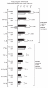
Cytokine expression elicited by S. typhimurium in SIV-infected (n = 4) and SIV-negative control macaques (n = 4) 5 h after infection. Fold changes in gene expression observed in a S. typhimurium–infected loop compared to a mock-infected loop from the same macaque were determined. Data are shown as geometric means of fold-increases ± s.e.m. Statistically significant (P < 0.05) differences are indicated. n.s., not significant.
Figure 3
Figure 3
Kinetics of lymphocyte depletion and viral replication in SIV-infected macaques compared to naive controls. (a) CD4+ T cell counts in peripheral blood of SIV-infected macaques (MK3, MK4, MK6 and MK10) over the time course of the experiment. (b) Viral loads in peripheral blood of SIV-infected macaques over the time course of the experiment. A dashed line indicates the limit of detection. (c) CD4+ and CD8+ T cell counts in peripheral blood of naive controls compared to SIV-infected macaques at the time of surgery. (d) Fraction of CD4+ T cells in the ileal lamina propria of naive controls compared to SIV-infected macaques at the time of surgery.
Figure 4
Figure 4
Analysis of lamina propria T lymphocytes. (a) T cells in the ileal lamina propria of SIV-infected macaques (n = 4) compared to naive controls (n = 6). The y-axis indicates the numbers of CD4+ or CD8+ T cells as percentage of the total number of lamina propria CD3+ Tcells. (b) Correlation between the fraction of CD4+CD8 T cells and the fold increases in IL-17 expression elicited in the ileal mucosa 5 h after S. typhimurium infection of SIV-infected macaques (n = 4) or SIV-negative macaques (n = 4). The y-axis indicates the numbers of CD4+ T cells as percentage of the total number of lamina propria CD3+ Tcells. (c,d) Cytokine expression by CD3+ lamina propria T cells from SIV-infected macaques (n = 4) or naive controls (n = 6) in response to PMA-ionomycin stimulation. (c) The y-axis indicates the numbers of IL-17–producing CD4+ or CD8+ T cells as percentage of the total number of lamina propria CD3+ Tcells. (d) The y-axis indicates the numbers of IFN-γ–producing CD4+ or CD8+ T cells as percentage of the total number of lamina propria CD3+ T cells. Bars represent geometric means ± s.e.m. Statistically significant (P < 0.05) differences are indicated.
Figure 5
Figure 5
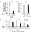
Bacterial translocation from the intestinal mucosa to internal organs in rhesus macaques and mice. (a) Recovery of S. typhimurium from the mesenteric lymph nodes of macaques 8 h after infection of ligated ileal loops. Data represent geometric means from control macaques or SIV-infected macaques ± s.e.m. (b,c) Recovery of S. typhimurium from the cecum (b) or internal organs (c) of C57BL/6 mice or Il17ra−/− mice 48 h after infection. Data represent geometric means ± s.e.m. Statistically significant (P < 0.05) differences are indicated.
Figure 6
Figure 6
Host responses elicited 48 h after S. typhimurium infection in the cecal mucosa of C57BL/6 mice orIl17ra−/− mice. (af) Data are expressed as fold changes of mRNA levels over mRNA levels detected in mock-infected C57BL/6 mice. Data represent geometric means ± s.e.m. Statistically significant (P < 0.05) differences are indicated. (g) Numbers of neutrophils per microscopic field (y-axis) were determined by a veterinary pathologist during a blinded examination of slides from the cecal mucosa. Data represent means ± s.e.m. Statistically significant (P < 0.05) differences are indicated.

"V体育官网入口" References

    1. Zhang S, et al. Molecular pathogenesis of Salmonella enterica serotype typhimurium–induced diarrhea. Infect. Immun. 2003;71:1–12. - PMC - PubMed
    1. Hohmann EL. Nontyphoidal salmonellosis. Clin. Infect. Dis. 2001;32:263–269. - PubMed
    1. Gordon MA, et al. Bacteraemia and mortality among adult medical admissions in Malawi-predominance of non-typhi salmonellae and Streptococcus pneumoniae. J. Infect. 2001;42:44–49. - "V体育安卓版" PubMed
    1. Alausa KO, et al. Septicaemia in the tropics. A prospective epidemiological study of 146 patients with a high case fatality rate. Scand. J. Infect. Dis. 1977;9:181–185. - PubMed
    1. Kankwatira AM, Mwafulirwa GA, Gordon MA. Non-typhoidal salmonella bacteraemia—an under-recognized feature of AIDS in African adults. Trop. Doct. 2004;34:198–200. - PubMed

"V体育2025版" Publication types

MeSH terms