V体育安卓版 - Skip to main page content
U.S. flag

An official website of the United States government

Dot gov

The VSports app下载. gov means it’s official. Federal government websites often end in . gov or . mil. Before sharing sensitive information, make sure you’re on a federal government site. .

Https

The site is secure V体育官网. The https:// ensures that you are connecting to the official website and that any information you provide is encrypted and transmitted securely. .

. 2008 Sep;125(1):91-100.
doi: 10.1111/j.1365-2567.2008.02824.x. Epub 2008 Feb 27.

Dexamethasone transforms lipopolysaccharide-stimulated human blood myeloid dendritic cells into myeloid dendritic cells that prime interleukin-10 production in T cells

Affiliations

Dexamethasone transforms lipopolysaccharide-stimulated human blood myeloid dendritic cells into myeloid dendritic cells that prime interleukin-10 production in T cells (V体育2025版)

Brenda M Bosma et al. Immunology. 2008 Sep.

Abstract

Myeloid dendritic cells (MDC) play an important role in antigen-specific immunity and tolerance. In transplantation setting donor-derived MDC are a promising tool to realize donor-specific tolerance. Current protocols enable generation of tolerogenic donor MDC from human monocytes during 1-week cultures. However, for clinical application in transplantation medicine, a rapidly available source of tolerogenic MDC is desired. In this study we investigated whether primary human blood MDC could be transformed into tolerogenic MDC using dexamethasone (dex) and lipopolysaccharide (LPS). Human blood MDC were cultured with dex and subsequently matured with LPS in the presence or absence of dex. Activation of MDC with LPS after pretreatment with dex did not prevent maturation into immunostimulatory MDC VSports手机版. In contrast, simultaneous treatment with dex and LPS yielded tolerogenic MDC, that had a reduced expression of CD86 and CD83, that poorly stimulated allogeneic T-cell proliferation and production of T helper 1 (Th1) cytokines, and primed production of the immunoregulatory cytokine interleukin-10 (IL-10) in T cells. In vitro, however, these tolerogenic MDC did not induce permanent donor-specific hyporesponsiveness in T cells. Importantly, tolerogenic MDC obtained by LPS stimulation in the presence of dex did not convert into immunostimulatory MDC after subsequent activation with different maturation stimuli. In conclusion, these findings demonstrate that combined treatment with dex and LPS transforms primary human blood MDC into tolerogenic MDC that are impaired to stimulate Th1 cytokines, but strongly prime the production of the immunoregulatory cytokine IL-10 in T cells, and are resistant to maturation stimuli. This strategy enables rapid generation of tolerogenic donor-derived MDC for immunotherapy in clinical transplantation. .


Figures
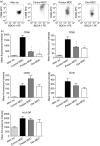
Figure 1
Figure 1
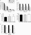
Effect of dex treatment on immunophenotype of human blood MDC. Control and predex MDC were generated by incubating isolated human blood MDC in the presence or absence of 1 μm dex for 18 hr and subsequently maturing them with 100 pg/ml LPS. Before addition of LPS the MDC were extensively washed to remove residual dex. Dex MDC were generated in the continuous presence 1 μm dex during both preincuation and LPS stimulation. (a) Representative dotplots of CD86 expression on freshly isolated (after iso), control, predex and dex MDC. (b) The immunophenotypic characteristics of freshly isolated (after iso), control, predex and dex MDC as analysed by flow cytometry. Depicted is the mean fluorescence intensity of the various markers. Data represent the means with SEM of 10 separate experiments. *P < 0·04, **P < 0·02 for the comparison of predex or dex MDC versus control MDC.
Figure 2
Figure 2
Continuous presence of dex during MDC maturation results in an impaired T-cell stimulatory capacity of MDC. Control, predex and dex MDC were generated by incubating isolated human blood MDC in the presence or absence of 1 μm dex for 18 hr and subsequently maturing them with 100 pg/ml LPS in the presence or absence of 1 μm dex for 24 hr. Before addition of allogeneic T cells, MDC were extensively washed to remove dex from the cultures. Allogeneic T-cell proliferation was assessed after 5 days of incubation with graded number of MDC by [3H]-thymidine incorporation. (a) A representative experiment showing T-cell proliferation induced by control, predex and dex MDC from the same donor. (b) Relative T-cell activation by predex and dex MDC compared to control MDC in eight experiments. To exclude variations in absolute proliferation between experiments, [3H]-thymidine incorporation in T cells stimulated with control MDC was normalized to 100% for each MDC number, thereby allowing easy pairwise comparison. Proliferation of T cells stimulated by predex and dex MDC was calculated by the following formula: Counts of T cells stimulated with predex or dex MDC divided by counts of T cells stimulated by control MDC times 100%. Data represent mean with SEM of eight separate experiments. *P < 0·01 for comparison of dex MDC versus control MDC. (c) Percentage recovery of control, predex, and dex MDC from wells after LPS activation. (d) Viability of MDC, determined by trypan blue exclusion, after LPS activation. (e) Relative T-cell activation by recounted dex MDC compared to recounted control MDC. MDC were recounted after LPS stimulation and graded numbers of MDC were used to stimulate allogeneic T cells. Data represent mean with SEM of five separate experiments.
Figure 3
Figure 3
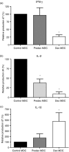
Dex MDC have reduced capacity to stimulate Th1 cytokine production but induce secretion of IL-10 by T-cells. (a) Relative IFN-γ production by predex and dex MDC compared to control-MDC on day 7. Supernatants of T cells stimulated by control MDC contained on average 456 ± 131 pg/ml IFN-γ. To exclude variations in absolute IFN-γ production between experiments, IFN-γ concentration in culture media of T cells stimulated with control MDC was normalized to 100%, thereby allowing easy pairwise comparison. Data represent mean with SEM of seven separate experiments. (b) Relative IL-2 production by control, predex and dex MDC on day 4. Supernatants of T cells stimulated by control MDC contained 260 ± 100 pg/ml IL-2. (c) Relative IL-10 production by control, predex and dex MDC on day 7. Supernatants of T cells stimulated by control MDC contained 10 ± 1·3 pg/ml IL-10. Data represent mean with SEM of six separate experiments. *P ≤ 0·03 for comparison of dex MDC versus control MDC.
Figure 4
Figure 4
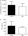
Upon restimulation dex MDC-primed T cells proliferate and produce IFN-γ comparable to control MDC-primed T cells. 15 × 104 allogeneic T cells were primed with 20 × 103 control or dex MDC. At day 7 the T cells were rested for 2 days in refreshed medium. T cells (75 × 103) primed with control or dex MDC were restimulated with 75 × 102 mature mo-MDC originating from the same donor as the control- and dex MDC. (a) T-cell proliferation was assessed by [3H]-thymidine incorporation 4 days after restimulation of the control or dex MDC primed T cells. (b) IFN-γ production in the supernatant on day 4 of restimulation was determined by ELISA. Data represent mean with SEM of three separate experiments.
Figure 5
Figure 5
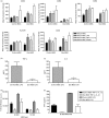
Dex MDC are functionally resistant to a second maturation stimulus in the absence of dex. Control- and dex MDC were exposed to a second maturation stimulus in the absence of dex consisting of either LPS (100 pg/ml), TNF-α + IL-1β (50 pg/ml) or CD40L (5 × 10³ 000 J558 cells) for 24 hr. (a) The immunophenotypic characteristics of these MDC were determined by flow cytometry. Depicted is the mean fluorescence intensity of the various markers on control and dex MDC before (mature MDC) and after the second stimulus (mature MDC LPS, mature MDC TNF-α + IL-1β, mature MDC CD40L). Data represent mean with SEM of three separate experiments. (b) TNF-α levels in the culture supernatants of control and dex MDC 24 hr after restimulation with LPS. Data represent the mean with SEM of six separate experiments. *P = 0·03 for the comparison of dex MDC versus control MDC. (c) IL-6 levels in the culture supernatants of control or dex MDC 24 hr after restimulation with LPS. Data represent the mean with SEM of six separate experiments. *P = 0·03 for the comparison of dex MDC versus control MDC. (d) Allogeneic T-cell proliferation primed by control or dex MDC from the same donor that had been restimulated with LPS or TNF-α + IL-1β (one representative experiment out of three). (e) IFN-γ production in the supernatant on day 5 of the T cells stimulated by control or dex MDC that had been restimulated with LPS or TNF-α + IL-1β. Data represent mean with SEM of three separate experiments.

References

    1. Banchereau J, Steinman RM. Dendritic cells and the control of immunity. Nature. 1998;392:245–52. - PubMed
    1. Fu F, Li Y, Qian S, Lu L, Chambers F, Starzl TE, Fung JJ, Thomson AW. Costimulatory molecule-deficient dendritic cell progenitors (MHC class II+, CD80dim, CD86–) prolong cardiac allograft survival in nonimmunosuppressed recipients. Transplantation. 1996;62:659–65. - PMC - PubMed
    1. Lutz MB, Suri RM, Niimi M, Ogilvie AL, Kukutsch NA, Rossner S, Schuler G, Austyn JM. Immature dendritic cells generated with low doses of GM-CSF in the absence of IL-4 are maturation resistant and prolong allograft survival in vivo. Eur J Immunol. 2000;30:1813–22. - PubMed
    1. Morelli AE, Thomson AW. Dendritic cells: regulators of alloimmunity and opportunities for tolerance induction. Immunol Rev. 2003;196:125–46. - PubMed (VSports注册入口)
    1. Boumpas DT, Chrousos GP, Wilder RL, Cupps TR, Balow JE. Glucocorticoid therapy for immune-mediated diseases: basic and clinical correlates. Ann Intern Med. 1993;119:1198–208. - PubMed

MeSH terms

VSports最新版本 - Substances

V体育官网 - LinkOut - more resources