Skip to main page content (V体育官网)
U.S. flag

An official website of the United States government

Dot gov

The . gov means it’s official. Federal government websites often end in VSports app下载. gov or . mil. Before sharing sensitive information, make sure you’re on a federal government site. .

Https

The site is secure. The https:// ensures that you are connecting to the official website and that any information you provide is encrypted and transmitted securely V体育官网. .

. 2008 Feb;13(2):141-52.
doi: 10.1016/j.ccr.2008.01.011.

GATA-3 links tumor differentiation and dissemination in a luminal breast cancer model

Affiliations

V体育安卓版 - GATA-3 links tumor differentiation and dissemination in a luminal breast cancer model

Hosein Kouros-Mehr et al. Cancer Cell. 2008 Feb.

Abstract

How breast cancers are able to disseminate and metastasize is poorly understood. Using a hyperplasia transplant system, we show that tumor dissemination and metastasis occur in discrete steps during tumor progression. Bioinformatic analysis revealed that loss of the transcription factor GATA-3 marked progression from adenoma to early carcinoma and onset of tumor dissemination. Restoration of GATA-3 in late carcinomas induced tumor differentiation and suppressed tumor dissemination VSports手机版. Targeted deletion of GATA-3 in early tumors led to apoptosis of differentiated cells, indicating that its loss is not sufficient for malignant conversion. Rather, malignant progression occurred with an expanding GATA-3-negative tumor cell population. These data indicate that GATA-3 regulates tumor differentiation and suppresses tumor dissemination in breast cancer. .


Figures
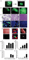
Figure 1
Figure 1. Dissemination and metastasis occurs at distinct progression steps in a breast cancer hyperplasia transplant model
(A) Focal hyperplasia isolated from a 3-week-old MMTV-PyMT; β-actin GFP mouse. (B–D) Representative (B) GFP whole mount, (C) H&E staining and (D) β–casein (green)/DAPI(blue) immunofluorescence of 5-, 8-, and 18-week tumor outgrowths. White arrowhead denotes early carcinoma adjacent to adenoma (yellow arrow) in an 8-week outgrowth. (E) Brightfield (upper) and GFP (lower) whole mount of the lungs of an 18-week tumor-bearing mouse. (F) Immunofluorescence of GFP (green), DAPI counterstain (blue) and tomato lectin (vasculature, red) of disseminated tumor cells in the lung of a tumor-bearing mouse (18-week outgrowth). (G) Brightfield (upper) and GFP (lower) whole mount of a lung with metastasis (arrowhead). Shown is the lung of a mouse sacrificed 8 weeks after removal of an 18-week tumor outgrowth. (H,I) Tumor dissemination rate (% mice with dissemination) (H), and frequency (total projected number of disseminated cells)(I) in the lungs of tumor-bearing mice at 5- (n=5), 8- (n=8), 12- (n=7), 15- (n=4), and 18-weeks (n= 7) post-transplant. (J,K) Lung metastasis rate (% mice with lung metastases) (J) and frequency (total number of lung metastases) (K) of tumor outgrowths. Mice with tumor outgrowths of 5 (n=5), 8 (n=5), 12 (n=5), 15 (n=6), and 18 weeks (n=5) underwent mastectomy and were sacrificed 8 weeks later. Data are represented as mean ± s.e.m. (I,K). Scale bars correspond to 1 mm (A, B, E, G), 25 µm (C) and 5 µm (D, F).
Figure 2
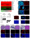
Figure 2. Loss of GATA-3 marks malignant progression in breast cancer
(A) Clustered image map of microarray data comparing Cy5-labelled (red) 5-week tumor outgrowth and Cy3-labelled (green) 18-week outgrowth (n = 3 microarrays). Color intensity represents M = log2 (Cy5 fluorescence / Cy3 fluorescence). ER, estrogen receptor; CA, carbonic anhydrase; WAP, whey acidic protein. (B) Immunofluorescence staining for GFP (green) and GATA-3 (red) and DAPI counterstain (blue) in 5-, 8-, and 18-week tumor outgrowths (n = 3 per condition). White arrow denotes early carcinoma in 8-week tumor outgrowth adjacent to an adenoma (yellow arrow). (C) Relative GATA-3 expression levels in nine breast cancer cell lines from a microarray dataset (Ross et al., 2000). Data are represented as log2 ratios of GATA-3 expression relative to control genes from a reference RNA in the pool of 60 cell lines. (D) Immunofluorescence staining for GATA-3 (red) and DAPI (blue) counterstain in a representative 18-week tumor outgrowth in which tumor dissemination to lungs was undetectable by histologic analysis. (E) Immunofluorescence staining of a representative disseminated cell in the lung of an 8-week tumor-bearing mouse (same color scheme as B). (F) Immunofluorescence staining for GATA-3 (red) and DAPI (blue) in a representative lung metastasis from a MMTV-PyMT mouse. (G–I) H&E staining and immunofluorescence for GATA-3 and DAPI in the MMTV-Wnt, MMTV-Neu, and C3(1)/Tag mouse models of breast cancer. Representative adenomas and carcinomas are shown from 9-month-old MMTV-Neu mice in the FVB/n strain (n = 12 mammary glands from 3 mice), 6-month-old C3(1)/Tag mice in the FVB/n strain (n =11 mammary glands from 3 mice), and 4.5-month-old MMTV-Wnt1 mice in the FVB/n strain (n = 8 mammary glands from 3 mice) that were analyzed for GATA-3 expression. Scale bars correspond to 25 µm (B, D, F–I) and 10 µm (E).
Figure 3
Figure 3. GATA-3 causally regulates tumor differentiation and dissemination in breast cancer
(A) H&E staining and immunocytochemistry for β-casein, perlecan (heparan sulfate proteoglycan), GFP, GATA-3, and nuclei (DAPI) in vector control and GATA-3 retrovirally transduced tumor outgrowths. Primary cultures of adenocarcinomas from 14-week-old MMTV-PyMT mice (non-fluorescent) were transduced with retrovirus, transplanted into the mammary fat pads of wild-type mice, and grown for 6 weeks. (B) Clustered image map of microarray data comparing Cy5-labelled (red) GATA-3 retrovirally-infected tumor outgrowths and Cy3-labelled (green) control-infected tumor outgrowths (n = 3 microarrays). Color intensity represents M = log2 (Cy5 fluorescence /Cy3 fluorescence). ER, estrogen receptor; CA, carbonic anhydrase. (C, D) Volume of tumor outgrowths and frequency of tumor dissemination in lungs of vector control- (n = 5) and GATA-3- (n = 5) infected tumor outgrowths. (E,F) Immunofluorescence for GATA-3 (red), GFP (green) and DAPI (blue) in (E) GATA-3-infected tumors and (F) in a disseminated tumor cell in lung arising from a GATA-3-infected tumor. Error bars indicate s.e.m.; *, P < 0.01 (Student’s t-test). Scale bars correspond to 25 µm (A, E), 10 µm (F).
Figure 4
Figure 4. Deletion of GATA-3 is not tolerated in differentiated tumors
(A,B) H&E staining and immunofluorescence for GATA-3, GFP as a marker of Cre-mediated deletion (CreGFP), β-casein as a differentiation marker, Keratin 18 as a luminal epithelial marker, a-smooth muscle actin (SMA) as a marker of myoepithelial cells, cleaved caspase-3 as a marker of apoptosis, and DAPI counterstain in tumors of 10-week-old MMTV-PyMT; WAP-rtTA-Cre; GATA-3fl/+ and MMTV-PyMT; WAP-rtTA-Cre; GATA-3fl/fl mice treated with doxycycline for (A) 2 weeks (n = 8 tumors) or (B) 8 weeks (n = 16 tumors). Black arrows indicate caspase-3 positive cells in the ductal lumen. Inserts are higher magnification views. Scale bars correspond to 25 µm. (C) Mean tumor volume of tumors at 8 weeks of doxycycline for MMTV-PyMT; WAP-rtTA-Cre; GATA-3fl/+ (GATA-3fl/+) and MMTV-PyMT; WAP-rtTA-Cre; GATA-3fl/fl (GATA-3fl/fl). Error bars indicate s.e.m. (n = 16 tumors); *, P< 0.01 (Student’s t-test).
Figure 5
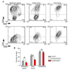
Figure 5. A stem cell-like population is enriched in late carcinomas of in MMTV-PyMT tumors
(A) CD24/CD29 and CD61/CD29 contour plots of C57BL/6 primary cells isolated from wild-type mammary glands, 11-week early, nonpalpable MMTV-PyMT tumor and a 5-month MMTV-PyMT tumor measuring 13×20×8mm (C57BL/6). Values in each quadrant indicate the percentage of cells in each condition (n = 10,000 events). (B) Percentage of CD61+, CD24+CD29+ double positive, and CD24+CD61+ double positive cells in samples described in (A). A total of 3–4 samples were analyzed per condition. Error bars indicate s.e.m.; *, P < 0.001 (Analysis of Variance).
Figure 6
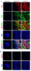
Figure 6. Immunohistochemical staining of CD29, CD61 and GATA-3 in MMTV-PyMT tumors
(A) Immunoflourescence for CD29 (green), CD61 (red), and DAPI (blue) on frozen sections of 7-week-old (early) and 14-week-old (late) MMTV-PyMT tumors. Early1 and Early2 refers to two distinct areas within the same tumor. (B) Immuflourescence for GATA-3 (red) and DAPI (blue) on serial sections of the tumors in (A). Adjacent, acetone-fixed frozen sections were thawed, processed with microwave antigen retrieval, and labelled with GATA-3 as described (Kouros-Mehr, 2006). Scale bar, 25 µm.
Figure 7
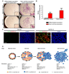
Figure 7. Stem cell-like populations from MMTV-PyMT tumors show increased motility and ability to differentiate into luminal and myoepithelial cells
(A, B) Analysis of motility among distinct cell populations purified from wild-type mammary glands and early MMTV-PyMT tumors. (A) Transwell migration assays performed on CD24+CD29+CD61+ (stem cell-like) and CD24+CD29CD61 (differentiated cell) subpopulations purified from normal mammary glands (C57BL/6, n = 2) and adenomatous MMTV-PyMT tumors (C57BL/6, n = 2). Cells were isolated with a FACSAria cell sorter and 1.5 × 10⁵ cells were used per condition. After 72 hours, (A) the membranes containing migratory cells were stained and (B) the fraction of the membranes containing stained cells were determined using Adobe Photoshop. Insets are higher magnifications of cells on the membranes. Scale bar, 0.5 mm. Error bars indicate s.e.m. (C) GATA-3 transduced tumor outgrowths have both luminal and myoepithelial cells. Primary cultures of adenocarcinomas from 14-week-old MMTV-PyMT mice (non-fluorescent) were transduced with retroviruses encoding vector alone (Vector) or GATA-3 (GATA-3), transplanted into the mammary fat pads of wild-type mice, grown for 6 weeks, and then stained for GATA-3 (red) and α-smooth-muscle-actin (SMA, green) by immunofluorescence, and nuclear staining with DAPI. Scale bar, 25 µm. (D) Model showing how GATA-3 expression and differentiated luminal epithelial markers may be lost through expansion of a GATA-3 negative stem cell-like cell population during MMTV-PyMT tumor progression. ER, estrogen receptor; WAP, whey acidic protein.

References (V体育官网入口)

    1. Asselin-Labat ML, Sutherland KD, Barker H, Thomas R, Shackleton M, Forrest NC, Hartley L, Robb L, Grosveld FG, van der Wees J, et al. Gata-3 is an essential regulator of mammary-gland morphogenesis and luminal-cell differentiation. Nat Cell Biol. 2007;9:201–209. - PubMed
    1. Barczak A, Rodriguez MW, Hanspers K, Koth LL, Tai YC, Bolstad BM, Speed TP, Erle DJ. Spotted long oligonucleotide arrays for human gene expression analysis. Genome Res. 2003;13:1775–1785. - PMC - PubMed
    1. Bertucci F, Houlgatte R, Benziane A, Granjeaud S, Adelaide J, Tagett R, Loriod B, Jacquemier J, Viens P, Jordan B, et al. Gene expression profiling of primary breast carcinomas using arrays of candidate genes. Hum Mol Genet. 2000;9:2981–2991. - PubMed
    1. Bloom HJ, Richardson WW. Histological grading and prognosis in breast cancer; a study of 1409 cases of which 359 have been followed for 15 years. Br J Cancer. 1957;11:359–377. - PMC (V体育官网入口) - PubMed
    1. Chambers AF, Groom AC, MacDonald IC. Dissemination and growth of cancer cells in metastatic sites. Nat Rev Cancer. 2002;2:563–572. - PubMed

Publication types

VSports - MeSH terms